Research Article | | Peer-Reviewed

Advances in Rapid Nucleic Acid Diagnostics for Disease Prevention and Control

Received: 23 April 2026     Accepted: 25 June 2026     Published: 6 August 2026
Views:       Downloads:
Abstract

Animal diseases present a major challenge to sustainable livestock production and global public health security. Rapid nucleic acid diagnostics, characterized by high specificity, sensitivity, and speed, have emerged as indispensable tools for early disease detection and precise control. This review provides a comprehensive overview of the recent advancements in rapid nucleic acid diagnostic technologies for animal diseases. We systematically categorize and evaluate current platforms, focusing on isothermal amplification techniques, CRISPR-based assays, and microfluidic micro-systems. Particular attention is given to how modern iterations have revolutionized the diagnostic process, pushing sensitivity toward the single-molecule realm and reducing turnaround times from hours to minutes. Furthermore, this paper analyzes the critical roles of these rapid-response systems in pandemic surveillance, vaccine efficacy assessment, and cross-border biosecurity risk management. Finally, we address current technical bottlenecks, such as sample preparation complexity and field-deployability constraints. We conclude that the integration of functional nanomaterials and artificial intelligence (AI) will be the driving force behind the next generation of intelligent, sample-in-answer-out, and field-deployable diagnostic tools, ultimately shaping the future of smart veterinary medicine.

Published in American Journal of Biomedical and Life Sciences (Volume 14, Issue 4)
DOI 10.11648/j.ajbls.20261404.12
Page(s) 61-75
Creative Commons

This is an Open Access article, distributed under the terms of the Creative Commons Attribution 4.0 International License (http://creativecommons.org/licenses/by/4.0/), which permits unrestricted use, distribution and reproduction in any medium or format, provided the original work is properly cited.

Copyright

Copyright © The Author(s), 2026. Published by Science Publishing Group

Keywords

Animal Diseases, Control and Prevention, Rapid Nucleic Acid Diagnostics

1. Introduction
The repercussions of animal disease outbreaks extend beyond compromised livestock production to encompass food safety, public health, and global trade. The limitations of conventional diagnostic techniques, such as pathological observation, culture, and serology, are becoming apparent: despite their proven specificity, these methods are often too cumbersome, slow, and insufficiently sensitive for the rapid and precise diagnostics needed in contemporary agriculture. It is these very limitations that have established nucleic acid testing as a pivotal research frontier, with its hallmarks of high sensitivity, accuracy, and specificity. The advent of real-time fluorescent PCR marked the beginning of a continuous evolution in molecular detection, progressing from qualitative to quantitative, from single-plex to multi-plex, and from laboratory-bound procedures to on-site rapid testing. Real-time PCR protocols, including SYBR Green I and TaqMan probe methods, have been instrumental in standardizing pathogen molecular diagnosis. DNA microarrays enabled high-throughput parallel gene sequence detection, providing crucial data for analyzing multiple pathogen lineages. Isothermal amplification techniques, such as Loop-Mediated Isothermal Amplification (LAMP), Recombinase Polymerase Amplification (RPA), and Nucleic Acid Sequence-Based Amplification (NASBA), simplified the amplification process by operating at a constant temperature, significantly enhancing the convenience of point-of-care testing. However, this convenience often comes with trade-offs. For instance, while LAMP is renowned for its robust amplification, designing specific primers is more complex than for PCR, and it can be more prone to non-specific amplification if conditions are not meticulously optimized. RPA, operating at lower temperatures, is exceptionally suited for field use but may face challenges in reagent stability and cost. Therefore, the choice between these techniques is not straightforward; it hinges on a balance between the need for extreme sensitivity, operational simplicity, and cost-effectiveness for a given application. More recently, the emerging CRISPR/Cas systems, which integrate nucleic acid recognition with signal amplification, have broken through the limitations of traditional methods in throughput and sensitivity, offering new avenues for portable and ultra-sensitive diagnosis of animal pathogens. This review aims to systematically summarize the development and application of the aforementioned rapid nucleic acid testing technologies in the prevention and control of animal diseases. It compares their differences in detection sensitivity, throughput, cost, and field-deployment capability, analyzes their potential value in multi-pathogen surveillance and targeted control strategies, and discusses future trends in their integration with cutting-edge technologies such as microfluidic chips, nanoprobes, and artificial intelligence. We hope this work will provide a valuable reference for innovative research and the industrial translation of molecular diagnostics for animal diseases.
2. Real-time Fluorescent Quantitative PCR (RT-PCR/qPCR)
Conventional PCR, despite being a core tool in DNA studies, lacks the capability for reliable quantification. Addressing this gap, real-time PCR emerged as a transformative breakthrough. The technique integrates fluorescent reporters into the PCR mixture, permitting the direct observation of amplification kinetics through fluorescence. This is achieved by correlating the accumulation of fluorescent signal with the amplification of the target DNA, which facilitates accurate qualitative and quantitative assessment. Thus, RT-PCR not only detects the presence of a target gene but also quantifies its initial copy number .
2.1. SYBR Green I Method (Fluorescent Dye)
SYBR Green I is a green-excitation fluorescent dye that targets and intercalates into the minor groove of double-stranded DNA (dsDNA). Its fluorescence intensifies upon binding. Consequently, as dsDNA PCR products accumulate during amplification, the overall fluorescence signal increases. This signal amplification facilitates both the detection and quantification of the target analyte (Figure 1).
SYBR Green I has become one of the most important techniques in molecular diagnosis because of its simplicity, low cost and high detection efficiency. It is now extensively applied in areas including the early diagnosis of animal diseases, clinical monitoring, and epidemiological research . By enabling real-time monitoring of fluorescence changes during amplification to achieve nucleic acid quantification, this technique provides an efficient tool for the rapid identification and control of infectious diseases. In the context of animal pathogen detection, the SYBR Green I method has been successfully applied to the rapid detection and quantification of a wide range of viruses . Zhang et al. developed a SYBR Green I-based real-time PCR (qPCR) method for detecting infectious laryngotracheitis virus (ILTV), employing primers targeting the gB gene and using a constructed recombinant plasmid as a positive control. The assay achieved a detection sensitivity of 3.34×10³ copies/μL, substantially outperforming conventional PCR, while demonstrating high repeatability with a coefficient of variation of 3.35%. In another application for equine viral infections, a duplex/triplex SYBR Green-based real-time RT-PCR was developed. This multiplex assay enables the simultaneous detection and differentiation of Equine Infectious Anemia Virus (EIAV), West Nile Virus (WNV), and Japanese Encephalitis Virus (JEV), offering a cost-effective solution for rapid differential diagnosis in clinical equine diagnostics and surveillance . The analytical performance of the SYBR Green I method is well-established across multiple studies. It is characterized by a precise linear correlation of Ct values with standard concentration, high sensitivity, powerful specificity, and consistent reproducibility. These proven capabilities thereby establish it as a dependable molecular methodology for rapid pathogen diagnostics and precise quantitative analysis. The SYBR Green I method operates by emitting fluorescence upon binding to double-stranded DNA, offering high sensitivity. The specificity of the amplicon can be further verified through melting curve analysis . However, a key limitation arises because the dye fluoresces upon contact with any dsDNA, making it difficult to discriminate between the specific target and non-specific byproducts like primer-dimers. Consequently, its specificity and reproducibility are generally lower than those of probe-based assays. Furthermore, the method is not suitable for multiplex detection. Therefore, while probe-based methods are preferred for high-precision and clinical quantitative diagnostics, the dye-based approach finds its primary utility in research, teaching, and assay development phases. Future research could focus on integrating this method with microfluidics, nanoprobes, and portable platforms to achieve high-throughput, on-site rapid molecular detection of animal pathogens, thereby providing robust support for the early warning and precise control of animal diseases.
Figure 1. Schematic principle of the SYBR Green I assay. SYBR Green I intercalates into double-stranded DNA (dsDNA) during the amplification process, resulting in an enhancement of fluorescence intensity that correlates with the accumulation of PCR products. This mechanism allows for real-time monitoring of the amplification progress. Quantitative analysis of the target nucleic acid is achieved based on the relationship between the threshold cycle ($C_t$) value and the initial template concentration. Furthermore, the specificity of the amplification and the purity of the products can be evaluated through melting curve analysis.
2.2. TaqMan Probe Method (Fluorescent Probe)
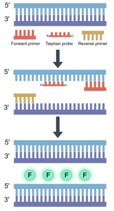
The TaqMan probe is a single-stranded oligonucleotide that is specifically complementary to the target DNA template. It is labeled with a fluorescent reporter dye at one end and a quencher moiety at the other. When the probe is intact, the fluorescence from the reporter is quenched due to its proximity to the quencher. During the PCR extension phase, when the polymerase reaches the probe bound to the template, its 5'→3' exonuclease activity cleaves the probe. This cleavage separates the reporter from the quencher, leading to the emission of a fluorescent signal. As the amplification products accumulate, the fluorescence signal increases exponentially, enabling real-time monitoring of the PCR process (Figure 2) . While it provides exceptional analytical specificity, sensitivity, and a low-background signal—properties that make it highly suitable for multiplexing, allelic discrimination, and precise quantification—the TaqMan method has notable limitations. The considerable expense and complexity associated with probe synthesis and optimization, combined with its vulnerability to target sequence variations, constitute a major bottleneck for applications like rapid variant pathogen detection or large-scale surveillance under budget constraints. The scalability of multiplexing is further limited by instrumental factors, namely the number of optical channels and spectral crosstalk.
TaqMan probe technology is now extensively utilized across multiple molecular detection fields, including human and animal pathogen identification, livestock and poultry quarantine, quality control of biological products, and vaccine potency evaluation. With continuous optimization of nucleic acid detection systems, research is increasingly focusing on achieving high-throughput, rapid, and sensitive pathogen detection through qPCR and multiplex PCR. For example, Dovas implemented TaqMan probe-based RT-PCR for high-throughput detection of H5N1 avian influenza virus in aquatic environments, while systematically investigating its survival characteristics and transmission patterns among different waterfowl species, providing crucial references for the eco-epidemiological study of waterfowl influenza. Min Zheng et al. designed specific primers and TaqMan MGB probes based on VP gene sequences, establishing a multiplex TaqMan MGB qPCR system for detecting various waterfowl parvoviruses (cGPV, MDPV, MDGPV, and SBDSV). This method demonstrated high specificity without cross-reactivity to other major waterfowl viruses, along with excellent repeatability and stability, enabling rapid, sensitive, and reliable differential detection of multiple parvoviruses and providing robust technical support for molecular diagnosis of waterfowl parvovirus infections . Furthermore, Liani Coronado et al. developed a dual-target multiplex RT-qPCR system based on TaqMan probes that integrates two validated PCR methods into a single reaction tube, enabling simultaneous identification of Classical Swine Fever Virus (CSFV) and African Swine Fever Virus (ASFV). This combined detection system showed no significant interference in analytical sensitivity for either target nucleic acid, maintained consistency with standard molecular methods, and exhibited excellent performance in clinical sample testing . Owing to their high throughput, strong specificity, and superior scalability, TaqMan probe-based multiplex qPCR and RT-qPCR technologies have become key molecular tools for animal pathogen nucleic acid testing, providing essential technical support for synchronous multi-pathogen detection, disease surveillance, and precision control.
Figure 2. Schematic principle of the TaqMan probe-based assay. The TaqMan probe is dual-labeled with a fluorescent reporter dye at the 5′ end and a quencher moiety at the 3′ end. During the extension phase of PCR, the 5′→3′ exonuclease activity of Taq DNA polymerase cleaves the hybridized probe, leading to the physical separation of the reporter from the quencher and the subsequent emission of a fluorescent signal. By monitoring the accumulation of this fluorescence, the target nucleic acid can be accurately quantified.
2.3. Rapid qPCR
Since its development in the early 1990s, probe-based quantitative PCR (qPCR) has become a cornerstone technology in molecular diagnostics and pathogen detection. Its evolution, progressing from the initial SYBR Green dye method to specific probe-based assays, enabled real-time monitoring of the amplification process, enhanced specificity, and reduced non-specific signal interference. However, as life science research advances into an era demanding rapid detection, high throughput, and field application, requirements for speed, specificity, and system simplicity have intensified. The limitations of traditional dye and probe methods—such as slow reaction speeds, complex setups, or high costs—have become increasingly apparent, thereby laying the groundwork for the emergence of "Rapid qPCR" technology.
In response to the growing demand for rapid testing, researchers began exploring ways to accelerate the reaction process without compromising high sensitivity and specificity. This endeavor marked a pivotal shift, leading to the conceptualization of Rapid qPCR. Rather than overhauling the fundamental principles of qPCR, this technology achieves a qualitative leap in detection efficiency through innovations at three key levels: system optimization, enzyme engineering, and thermal cycling acceleration . Rapid qPCR employs thermostable DNA polymerases and reverse transcriptases with superior reaction kinetics to minimize the duration of each amplification cycle . Technologically, it supersedes conventional metal blocks with advanced microfluidic chips and highly thermally conductive miniature heating systems, enabling temperature cycling within seconds . Kang et al. developed an ultra-fast chip-based real-time PCR system utilizing nanoplasmonic heating, where efficient photothermal conversion via gold nanostructures achieves millisecond-level thermal cycling, drastically reducing amplification time. This approach maintains sensitivity and accuracy comparable to conventional qPCR while completing detection in mere minutes . Furthermore, reducing the reaction volume (typically ≤10 μL) diminishes thermal inertia, thereby accelerating heat transfer . Nunez-Balto created a disposable silicon-based integrated micro-qPCR chip that consolidates sample processing and nucleic acid amplification. This system features rapid heating/cooling rates and efficient heat transfer characteristics, enabling rapid quantitative detection of pathogen nucleic acids. Combining high-performance optical components with sensitive detectors and real-time algorithm optimization enables reliable ultra-rapid signal reading. Ahn, J. S. developed a representative system leveraging the photothermal properties of gold nanoshells for instantaneous heating, dramatically speeding up the qPCR process. This method achieves high-sensitive detection in just minutes, paving the way for portable, field-deployable molecular diagnostics.
Rapid qPCR achieves not only a leap in speed but also a significant enhancement in overall performance compared to traditional systems. Crucially, it maintains the high specificity and sensitivity of probe-based chemistry, effectively avoiding the non-specific amplification interference common in dye-based methods. Meanwhile, optimized reaction buffers and enzyme systems improve amplification efficiency, ensuring stable Ct values and strong reproducibility even under shortened cycling conditions. In assay design, Rapid qPCR typically employs shorter amplicons (80–150 bp), which further accelerates cycling and improves amplification accuracy. Moreover, the technology offers a higher degree of integration: by combining sample preparation, nucleic acid extraction, amplification, and detection into a single system, it enables "sample-to-answer" workflows to be completed within just minutes . These characteristics make it particularly suitable for on-site infectious disease screening, emergency response during public health crises, and the development of portable molecular diagnostic devices.
Rapid qPCR offers a decisive speed advantage over conventional fluorescent methods, compressing the 60–120 minute workflow into a mere 10–30 minutes while preserving high analytical performance. The commercial availability of specialized reagents is steadily lowering the cost per test. Crucially, its independence from large instruments facilitates true point-of-care testing, driving the technology toward system integration and portability. This leap in velocity, coupled with emerging advancements in microfluidics, nanophotothermal materials, and AI, heralds a future where precise nucleic acid testing can be completed within minutes—a capability with profound implications for clinical diagnosis, public health, and precision medicine. Therefore, this technological progression signifies more than an incremental upgrade; it embodies a historic transition for molecular diagnostics from the centralized laboratory to the point-of-need, and from hour-scale to minute-scale operation.
3. DNA Microarray
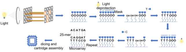
A DNA microarray (or gene chip) is a high-throughput technology that operates on nucleic acid hybridization. The method works by fixing thousands of unique DNA probes to a solid surface like glass or silicon. When a sample is applied, its nucleic acids bind specifically to complementary probes, allowing for the parallel measurement of multiple target sequences or their expression profiles . Leveraging high sensitivity and parallel analysis, DNA microarrays are widely used in gene expression studies, genomics, and infectious disease diagnostics (Figure 3) . Early research primarily focused on molecular identification and lineage analysis of pathogens. In 2006, Li et al. pioneered the use of a serotype-specific DNA microarray for the simultaneous detection of Shigella and pathogenic E. coli, achieving a sensitivity of 10⁴ CFU/mL in clinical stool samples . Subsequently, Maynard et al. demonstrated the detection of Salmonella enterica serovar Typhi in complex environmental samples, such as wastewater, by integrating PCR with microarray hybridization, with a detection sensitivity of approximately 0.1% of the total DNA . In the field of animal disease diagnostics, DNA microarray technology similarly exhibits broad application prospects. Xia et al. developed a multiplex detection system coupling quadruplex PCR amplification with a gene microarray readout, enabling the simultaneous detection and differentiation of various porcine viruses, including CSFV, African Swine Fever Virus (ASFV), and Atypical Porcine Pestivirus (APPV). Furthermore, this platform can distinguish vaccine strains from wild-type viruses, demonstrating excellent typing accuracy and providing a new high-throughput tool for precise diagnosis and epidemiological tracing of swine viral infections . Meanwhile, Gwida et al. applied DNA microarray technology to the systematic analysis of antimicrobial resistance and virulence gene profiles in E. coli of livestock origin. They constructed an oligonucleotide probe array encompassing multiple common resistance and virulence gene loci. This system allows for the concurrent detection of numerous resistance and virulence genes, offering an efficient molecular monitoring tool for assessing the risk of antimicrobial resistance transmission and the pathogenic potential of livestock-derived E. coli.
Figure 3. Schematic representation of the DNA microarray principle. A large number of oligonucleotide probes with known sequences are immobilized on the solid surface of the microarray. Fluorescently labeled target nucleic acids from the sample hybridize with these complementary probes. By scanning the signal intensity of each microdot (feature), parallel quantitative analysis of multiple targets can be achieved. This technology is characterized by high-throughput and massive parallelism, making it suitable for pathogen detection, genotyping, and gene expression profiling.
The distinguishing asset of DNA microarrays compared to conventional assays is their unparalleled capacity for multi-analyte detection. Evolving research, shaped by market needs, is now pivoting toward a deep convergence with nucleic acid amplification strategies, notably isothermal methods . This convergence addresses key limitations by simultaneously elevating sensitivity, accelerating throughput, and enabling true field-deployable (on-site) diagnostics. This is exemplified by microfluidic chips that host PCR or LAMP reactions for automated, parallel detection of common porcine pathogens, delivering integrated solutions to the swine industry A sophisticated manifestation of this trend is the nanoplasmonic microarray hybridized with solid-phase RPA, presented by Lee Their methodology anchors amplification via biotin-streptavidin binding and fine-tunes conditions to maximize surface plasmon-enhanced fluorescence (SPEF), yielding an exceptional sensitivity of 4 copies/reaction within 30 minutes—showcasing its potential for high-performance multi-target pathogen detection, including emerging threats like SARS-CoV-2. Parallel to hardware-centric systems, the emergence of paper-based biochips dramatically broadens the operational scope of DNA sensing. Hu et al. demonstrated this by executing LAMP directly on a paper chip with a smartphone as the detector, successfully identifying pathogens like E. coli O157: H7, Salmonella spp., and S. aureus at the point of need. The simplicity and ultra-low cost of this system make it a transformative tool for rapid screening in resource-constrained settings .
Since its emergence in the late 1990s, DNA microarray technology has undergone a significant evolution from low-density oligonucleotide arrays to high-density whole-genome chips, progressively achieving multi-target, rapid, and high-throughput nucleic acid detection. In animal disease diagnostics, microarrays enable the simultaneous detection of numerous pathogens and tens of thousands of genes or biomolecules at once. They offer the advantages of high throughput, high sensitivity (capable of detecting low-abundance targets), and compatibility with diverse samples, thereby enhancing diagnostic efficiency and playing a pivotal role in genotyping, mutation detection, and epidemiological research. The process is rapid and highly automated, significantly boosting analytical efficiency. Furthermore, the technology reduces reagent consumption and lowers costs, making it suitable for large-scale studies in gene expression profiling, genomic comparison, and mutation screening. However, sample preparation and labeling procedures for microarrays are relatively complex, involving multiple steps such as RNA/DNA extraction, transcription, and labeling, and require considerable technical expertise from operators. The massive datasets generated necessitate bioinformatics tools for interpretation, and probe non-specific binding can lead to false-positive signals. The high cost of high-resolution imaging equipment also limits its widespread adoption. In recent years, to overcome these limitations, researchers have integrated DNA microarrays with novel rapid detection technologies such as isothermal amplification (RPA, LAMP), CRISPR/Cas systems, and microfluidic platforms. This has created integrated "amplification-recognition-chip readout" systems that significantly improve sensitivity, specificity, and field applicability. The further integration of DNA microarrays with artificial intelligence and machine learning can enhance the accuracy and efficiency of data analysis, facilitating disease prediction, early diagnosis, and the development of personalized treatment strategies. With advancements in nanotechnology, the future of DNA microarrays lies in miniaturization and portability, deeper integration with novel signal amplification and automated detection technologies, enhanced parallel detection capabilities for multiple targets and samples, and broader application in the field-based rapid detection and monitoring of animal diseases. Through these technological convergences, DNA microarrays are poised to transition from a laboratory research tool to a practical, field-deployable diagnostic platform, enabling precise, rapid, and high-throughput prevention and control of animal diseases.
4. Isothermal Amplification Technology
Isothermal amplification technology allows for the efficient amplification of nucleic acids at a single, constant temperature. This process bypasses the need for thermal cycling, enabling real-time fluorescence detection. Unlike traditional PCR, it provides the benefits of simple instrumentation, faster results, and easier operation, without compromising sensitivity or specificity. This combination of features makes it ideally suited for rapid, field-deployable diagnostics in areas with limited resources.
4.1. LAMP
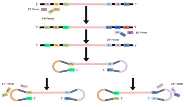
LAMP is a highly efficient nucleic acid amplification technique for in vitro applications, first pioneered by Notomi et al. in 2000 . It has garnered significant attention due to its operational simplicity and high amplification efficiency. The technique can achieve up to 10⁹-fold amplification of the target sequence within 60 minutes, offering high specificity, sensitivity, and low cost. Its mechanism hinges on six distinct regions at the 3' and 5' ends of the target gene, for which four specific primers (inner and outer primers for both upstream and downstream regions) are designed. The reaction system includes the template, the aforementioned primers, and a strand-displacing DNA polymerase. Under constant temperatures of 60–65°C, the template first forms a dumbbell-like structure facilitated by the enzyme and primers, followed by self-cycling amplification that generates a large quantity of product. The byproduct magnesium pyrophosphate precipitates in an amount proportional to the DNA yield, enabling result interpretation via turbidity measurement, colorimetric reaction, or gel electrophoresis (Figure 4) .
Figure 4. Schematic principle of the Loop-mediated Isothermal Amplification (LAMP) technique. The LAMP technique facilitates exponential amplification of target sequences at a constant temperature (60-65°C) using a strand-displacement DNA polymerase and a set of primers that recognize six distinct regions on the target. This process leads to the formation of characteristic loop structures and cauliflower-like DNA concatemers. The resulting magnesium pyrophosphate precipitate or changes in fluorescent signals can be interpreted via visual inspection or instrumental detection, enabling rapid and sensitive diagnosis.
Building on this foundation, Stratakos developed a multiplex LAMP (M-LAMP) system capable of the simultaneous quantitative detection and differential diagnosis of E. coli and enterotoxigenic E. coli in beef and fecal samples. Utilizing RT-LAMP, Salmonella and Vibrio parahaemolyticus could be distinguished within 60 minutes in a mixture based on differences in their Tm values . Furthermore, the technique has been successfully applied to the specific amplification of H5 subtype viral RNA from extracted viral RNA encompassing H1-H15 hemagglutinin subtypes of avian influenza virus and human pathogenic respiratory viruses . For the detection of influenza A (H1, H3) and influenza B viruses, the method demonstrated single-genome-copy sensitivity and completed diagnosis within 40 minutes, even without prior RNA extraction . The comparative analysis by Chen demonstrated the superior sensitivity of RT-LAMP over RT-PCR in the preclinical detection of CSFV. In a distinct application, Xu et al. devised a LAMP assay targeting the T. cruzi HSP70 gene, capable of specific amplification across diverse genetic lineages within one hour. This assay, notable for its long-term stability at -20°C and design prioritizing accessibility, addresses critical needs for Chagas disease surveillance in low-resource regions . Advancing multiplex capabilities, a triplex system integrating LAMP with a lateral flow dipstick (LFD) was established for the concurrent detection of BVDV, BRV, and BPV, delivering results in about 30 minutes with a detection limit of 2.4×10¹ copies/μL . Complementing these biochemical advancements, Cho J et al. created a portable, modular RT-LAMP device for on-site detection of Viral Hemorrhagic Septicemia Virus (VHSV) in aquaculture. This integrated hardware solution, which merges heating and fluidic control, enables rapid, point-of-care diagnosis, signifying a crucial advance for disease management in remote aquaculture operations with limited infrastructure .
The LAMP technique is characterized by high amplification efficiency, excellent sensitivity, strong specificity, and operational simplicity. The reaction is conducted at 60–65°C, and its products can be detected through rapid visual readouts such as fluorescence, turbidity, or color change, making it particularly suitable for pathogen monitoring in field settings and resource-limited environments. Nonetheless, the technology presents certain challenges, including complex primer design, susceptibility to amplicon contamination, difficulties in achieving multiplex detection, and limited capabilities for quantitative analysis. In recent years, the integration of LAMP with microfluidic chips, CRISPR/Cas systems, and portable detection devices has demonstrated a promising future for its application in rapid, low-cost pathogen testing.
4.2. RPA
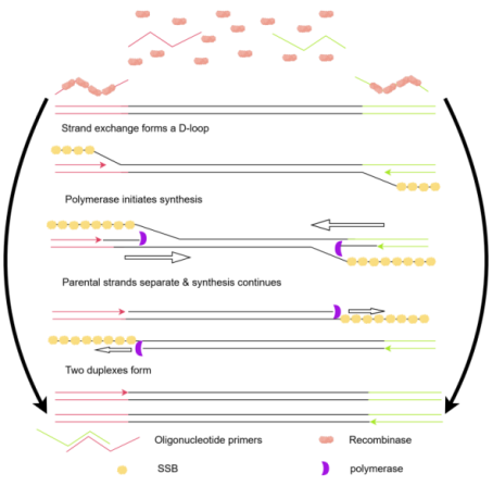
RPA was pioneered in 2006 by the UK-based company TwistDx . It is a rapid nucleic acid amplification method mediated by recombinase. The RPA reaction system comprises three core components: a recombinase (which binds to oligonucleotide primers), a single-stranded DNA-binding protein (SSB), and a strand-displacing DNA polymerase. Its optimal reaction temperature is 37 °C, and amplification is typically completed within 20 minutes . The recombinase-primer complexes specifically recognize the target double-stranded DNA, initiating a strand exchange reaction to start DNA synthesis. Catalyzed by the strand-displacing DNA polymerase, the target sequence is amplified exponentially, while the SSB stabilizes the displaced DNA strands to prevent non-specific binding (Figure 5).
Figure 5. Schematic workflow of the Recombinase Polymerase Amplification (RPA) process. RPA achieves rapid exponential amplification at a constant, low temperature (37-42°C). The process is initiated by the binding of a recombinase-primer complex to the target sequence, followed by DNA synthesis facilitated by the synergistic action of a strand-displacement DNA polymerase and single-stranded binding (SSB) proteins. The amplification products can be rapidly detected and visualized through either fluorogenic probes or lateral flow dipsticks (LFD).
In tuberculosis diagnostics, Boyle demonstrated that RPA exhibited significantly superior sensitivity and specificity compared to indirect fluorescence microscopy for detecting Mycobacterium tuberculosis. Xia developed a real-time RPA method that achieved a detection limit of 10 copies with 95% reproducibility. For veterinary applications, an RPA combined with a lateral flow dipstick (RPA-LFD) enabled rapid, visual detection of Newcastle disease virus and infectious bronchitis virus . In malaria diagnosis within resource-limited settings, an RPA-LFD system detected Plasmodium falciparum in just 15 minutes. Furthermore, Wu et al. showed that their assay could detect five different subtypes of human Bocavirus B without cross-reactivity to other subtypes or common respiratory pathogens . Separately, Zhu et al. established a simple and cost-effective real-time RPA assay for Feline Calicivirus (FCV), achieving a detection limit of 100 copies/μL in clinical samples. This method provides a practical alternative for the rapid and sensitive detection of FCV in laboratories and veterinary clinics, particularly those with limited facilities .
The practical appeal of RPA lies in its independence from sophisticated nucleic acid purification. Reaction products are compatible with diverse detection formats, from real-time monitoring with fluorescent probes to endpoint analysis using lateral flow dipsticks, biochips, or gel electrophoresis . These characteristics position it as a strong candidate for point-of-care testing in resource-constrained environments, dramatically reducing dependency on instrumentation while combining user-friendliness, specificity, and cost-effectiveness . The technique, however, is not without its limitations. Challenges encompass complicated primer design, constrained specificity, occasional reaction instability, and a propensity for false positives, driving current research to overcome hurdles in multiplexing and reliable quantification. Even so, RPA's core attributes underscore its substantial promise for developing rapid and low-power nucleic acid tests .
4.3. NASBA
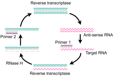
NASBA is an isothermal enzymatic technique specifically designed for amplifying RNA targets, such as 16S rRNA or mRNA, which allows for differentiating live from dead bacteria (Figure 6). This isothermal reaction employs two primers, AMV-RT, T7 RNA polymerase, and RNase H. Conducted at a constant 42 °C without thermal cycling, it can amplify RNA by 109–1012-fold within 2 hours and has been used for detecting various pathogens . For instance, a real-time NASBA assay for SARS-CoV-2 achieved a detection limit of 200 copies/mL with high clinical accuracy (97.64% PPA, 100% NPA) . Another study integrated NASBA with the CRISPR-Cas12a system to construct a "NASBA-Cas12a" detection platform for the rapid identification of Rotavirus A, a common pathogen causing childhood diarrhea. This method can be completed in approximately 70 minutes under isothermal conditions (37 °C), achieving a detection limit of about 1.2 copies/µL with high sensitivity and specificity, highlighting its potential for clinical rapid diagnostics .
The position of NASBA as a core technology for RNA pathogen detection is built upon a foundation of high sensitivity, real-time fluorescence capability, and procedural simplicity, delivering rapid and specific assays. Nonetheless, the technology has limitations: its system is relatively complex, demands high enzyme activity and stringent reaction conditions, exhibits poor product stability, and is exclusively applicable to RNA targets, which constrains its universal application. In recent years, the integration of NASBA with microfluidic chips and CRISPR/Cas detection platforms has opened new avenues for its development in the field of portable molecular diagnostics.
Figure 6. Schematic illustration of Nucleic Acid Sequence-Based Amplification (NASBA). The NASBA process begins with the reverse transcription of the target RNA into cDNA. Upon the degradation of the original RNA strand by RNase H, a second primer initiates a T7 RNA polymerase-mediated cyclic transcription process, leading to the continuous production of antisense RNA amplicons. This enzyme-driven cycle enables highly sensitive detection of RNA-based pathogens under isothermal conditions.
5. Nucleic Acid Detection Technology Based on CRISPR/Cas
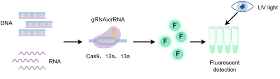
The The CRISPR/Cas system is an adaptive immune defense mechanism originating from bacteria and archaea, which functions to recognize and resist the invasion of foreign nucleic acids. Guided by a guide RNA, the Cas protein specifically recognizes the target nucleic acid sequence through base complementary pairing and exerts nuclease activity for cleavage (Figure 7) . The CRISPR/Cas9 system, renowned for its precise gene-editing capability, has become a revolutionary tool in genetic engineering. In recent years, various novel CRISPR/Cas systems have been successively discovered and widely applied in life science and medical research. Benefiting from the high sensitivity and specificity of the CRISPR system, multiple effector proteins (such as Cas9, Cas12a, and Cas13a/b) have been developed for molecular diagnostics. Nucleic acid detection technologies based on CRISPR show great potential in fields like point-of-care testing and field-based detection .
Figure 7. Schematic illustration of the mechanism of CRISPR/Cas systems. The CRISPR/Cas system achieves sequence-specific recognition and cleavage guided by crRNA. Specifically, Cas9 facilitates double-stranded DNA (dsDNA) cleavage under the guidance of a single-guide RNA (sgRNA). Cas12a activates non-specific trans-cleavage of single-stranded DNA (ssDNA) upon binding to its target DNA. Similarly, Cas13a triggers the degradation of collateral RNA molecules following the recognition of target RNA. These unique enzymatic properties have been leveraged to develop high-sensitivity nucleic acid detection platforms, such as DETECTR and SHERLOCK.
The foundational Cas9 protein from Type II CRISPR systems mediates precise, gRNA-directed cleavage of dsDNA. Its dual nuclease domains generate blunt-end breaks, effectively halting PCR amplification. This functionality can be strategically modulated: single-domain mutation produces a nickase (nCas9), and double mutation abolishes cleavage activity (dCas9) . The integration of the CRISPR/Cas9 system, with its specific enzymatic cleavage activity, with NASBA enables pathogen genotyping and SNP detection. A technology developed by Pardee could be lyophilized on paper strips and was successfully applied for the single-base resolution detection of Zika virus (ZIKV) in macaque plasma, serving as a reliable solution for point-of-care testing (POCT). Furthermore, the Cas-EXPAR method developed by Huang exhibits high detection sensitivity and can accurately distinguish single-base mismatches. This technique leverages CRISPR/Cas9 to perform specific cleavage, generating DNA fragments. These cleavage products then act as primers to initiate an Exponential Amplification Reaction (EXPAR), and the resulting amplicons are detected in real-time by binding with fluorescent dyes.
Cas12a, classified under the Type V-A CRISPR-Cas system, specifically recognizes target double-stranded DNA (dsDNA) guided by a crRNA and exhibits collateral nonspecific ssDNA cleavage activity. Upon the formation of the Cas12a/crRNA/target sequence ternary complex, Cas12a becomes activated and demonstrates DNase activity towards single-stranded DNA . The DETECTR system, developed in 2018, achieved rapid detection of human papillomavirus by integrating recombinase polymerase amplification (RPA) with the trans-cleavage activity of Cas12a . Furthermore, Wang established a method combining RPA and Cas12a cleavage within a single reaction system. This assay enables visual detection under blue light within 30 minutes, achieving single-molecule sensitivity and 100% accuracy for Mycoplasma detection, significantly outperforming conventional PCR methods.
Cas13a, a CRISPR effector that targets and cleaves RNA via crRNA guidance, exhibits collateral activity upon target recognition, indiscriminately degrading nearby RNA . The SHERLOCK platform, developed by Gootenberg , leverages CRISPR/Cas13a to achieve single-nucleotide specificity and has been successfully applied to distinguish Zika from Dengue virus serotypes, differentiate pathogen from host DNA, and identify cancer-associated mutations. The subsequently discovered Cas13b (Type VI-B) also exhibits trans RNA cleavage activity, and the optimized SHERLOCKv2 system further enhances detection sensitivity . In terms of technological application, Qin integrated Cas13a with a microfluidic chip to create a portable POCT system, which provides results via a miniaturized fluorometer within 5 minutes, facilitating field deployment. In the veterinary field, the RPA-Cas13a method established by Chang for detecting Porcine Reproductive and Respiratory Syndrome Virus (PRRSV) demonstrates high sensitivity and strong specificity, with no cross-reactivity against other common porcine viruses (PCV, PPV, CSFV, PRV), supporting both real-time fluorescence and visual readout. Platforms like SHERLOCK and DETECTR achieve attomolar-level sensitivity, while their integration with microfluidics and nanotechnology further enhances the practicality of on-site testing .
The significant promise of CRISPR-based nucleic acid detection lies in its operational ease, exceptional single-base specificity, high sensitivity, and cost-effectiveness. These attributes, combined with versatile readout methods, make it a compelling technology for rapid point-of-care applications across disease control, food safety, and environmental surveillance. Nevertheless, its translation faces practical constraints. The performance of Cas proteins is highly dependent on specific reaction parameters, and the considerable variation in optimal conditions among different systems constitutes a bottleneck for standardization and scaled manufacturing. A fundamental limitation is the nearly ubiquitous requirement for pre-amplification (e.g., via RPA or LAMP), which introduces amplification artifacts, elevates contamination risk, and inherently limits quantitative precision. Moreover, the stability of the core components (Cas ribonucleoproteins and crRNA) during storage, coupled with cost considerations, presents tangible barriers to deployment in grassroots or remote field settings.
In summary, CRISPR technology provides a highly specific, programmable, and portable strategy for rapid nucleic acid testing, demonstrating considerable advantages in disease diagnosis, pathogen tracking, and genetic mutation screening. Future efforts should prioritize the development of Cas proteins with enhanced thermal stability and faster reaction kinetics, the construction of integrated "sample-to-answer" detection systems, and the combination of CRISPR assays with microfluidic chips and intelligent signal readout platforms. These advancements will accelerate its translation into clinical diagnostics and field-deployment scenarios.
6. Summary and Application Prospects of Rapid Nucleic Acid Diagnostics
Rapid nucleic acid testing is revolutionizing the early diagnosis and control of animal diseases. Moving beyond conventional PCR to high-throughput DNA microarrays, and further to isothermal amplification and CRISPR/Cas systems, diagnostics are becoming more sensitive, simple, integrated, and field-ready. Many of these tools have already moved from the lab to clinical and field settings, greatly improving the speed and accuracy of animal pathogen surveillance. Fast, precise diagnosis is pivotal for early intervention and cutting transmission chains—especially for emerging and re-emerging infections. In agriculture, nucleic acid-based testing also proves equally promising: its high-throughput capacity enables early warning systems for crop diseases.
Overall, quantitative real-time PCR (qPCR) remains the “gold standard” for nucleic acid detection of animal diseases, owing to its high sensitivity, accurate quantification, and good reproducibility. In particular, the TaqMan probe–based approach has demonstrated outstanding performance in multiplex detection and clinical diagnostics. However, its strong dependence on sophisticated instrumentation and relatively time-consuming workflows limits its applicability for rapid on-site testing. By virtue of its high-throughput capacity and simultaneous multi-target detection, DNA microarray technology offers unique strengths in pathogen identification, molecular typing, and drug resistance surveillance. Nevertheless, its adoption is hampered by intricate sample processing, prohibitive costs, and the requirement for sophisticated instrumentation, confining its current utility to specialized research environments. By comparison, isothermal amplification technologies (e.g., LAMP, RPA, and NASBA) simplify procedures by operating at a uniform temperature. Specifically, LAMP excels in amplification efficiency and ease of visual readout, whereas RPA is more adaptable to field settings given its lower thermal requirements and speed. NASBA, meanwhile, maintains a distinctive edge in detecting RNA-based pathogens. However, common drawbacks such as intricate primer design, off-target amplification, and poor quantification persist. The emerging CRISPR/Cas platform has revolutionized the field by enhancing specificity and sensitivity via Cas12a/Cas13a-mediated trans-cleavage. Although it offers single-nucleotide resolution and diverse readout formats suitable for decentralized testing, its widespread adoption is still constrained by the necessity of initial amplification and the need for improved system robustness and standardization.
Current rapid nucleic acid diagnostics face challenges in effectively controlling precision and reliability. In CRISPR systems, the non-specific cleavage activity of Cas12a can readily lead to false-positive results. Inhibitors present in animal clinical samples also significantly compromise detection sensitivity. For isothermal amplification techniques, improper primer design may induce primer-dimer artifacts, posing a prominent risk of false positives, particularly when using fluorescent dye-based detection. To enhance precision, AI algorithms can be employed to predict gRNA secondary structures, thereby strengthening the specificity of gRNA binding to its target, alongside developing reagents resistant to matrix interference. Furthermore, constructing multiplex CRISPR detection systems by combining Cas13a and Cas12a enables simultaneous screening for mixed pathogens. Automated, intelligent primer design tools can generate highly efficient primer sets automatically, while engineering robust enzymes to improve their tolerance to sample inhibitors and adopting sealed microfluidic systems to prevent aerosol contamination will significantly enhance assay stability. On the front of standardization and industrialization, it is imperative to establish a unified set of detection protocols with clearly defined core metrics such as sensitivity and cross-reactivity. Promoting the miniaturization of devices will make result interpretation more convenient and efficient, and extending reagent shelf life will ensure a stable supply for remote areas.
To address these technical and dissemination challenges, a phased approach is recommended. Short-term efforts should prioritize minimizing CRISPR off-target effects while concurrently focusing on reducing the cost of RPA lateral flow strips. In the medium term, the goal should be establishing multiplexed microfluidic detection platforms for major livestock and poultry diseases. The long-term objective is to construct an intelligent biosecurity network integrating "mobile detection, cloud-based analysis, and real-time alerting," fostered through industry-academia-research collaboration. This requires dedicated efforts towards developing domestic high-performance enzyme reagents and implementing systematic training for grassroots-level testing personnel. Ultimately, these initiatives will facilitate a paradigm shift in disease control from passive treatment to active prevention. In summary, propelled by performance breakthroughs and scenario adaptability, rapid nucleic acid testing technologies have become a cornerstone of the infectious disease prevention and control system. The continued convergence with disciplines like synthetic biology and artificial intelligence will steer the field towards more intelligent and integrated solutions, thereby providing stronger safeguards for both human and animal health.
Abbreviations

LAMP

Loop-Mediated Isothermal Amplification

RPA

Recombinase Polymerase Amplification

NASBA

Nucleic Acid Sequence-Based Amplification

RT-PCR/qPCR

Real-time Fluorescent Quantitative PCR

dsDNA

double-stranded DNA

Author Contributions
Jinhui Sun: Conceptualization, Data curation, Writing – original draft
Baosheng Sun: Resources, Writing – review & editing
Xiuli Ruan: Project administration
Yinzhen Sheng: Funding acquisition, Supervision, Writing – review & editing
Funding
This work is supported by Guizhou Provincial Department of Agriculture and Rural Affairs (grant number GZMFCYJSCXTD-202603 and SXMZ-2026-05) and Guizhou Provincial Education Department (grant number Qian Jiao Han(2024)No. 44). The APC was funded by The Science and Technology Bureau of Zunyi City.
Data Availability Statement
The data supporting the outcome of this research work has been reported in this manuscript.
Conflicts of Interest
The authors declare no conflicts of interest.
References
[1] Freeman WM, Walker SJ, Vrana KE. Quantitative RT-PCR: pitfalls and potential. Biotechniques 26(1): 112-125.
[2] Silva SJRD, Kohl A, Pena L, Pardee K. Clinical and laboratory diagnosis of monkeypox (mpox): Current status and future directions. Iscience 26(6): 106759.
[3] Loor-Giler A, Castillo-Reyes S, Santander-Parra S, Caza M, Kyriakidis NC, Ferreira AJP, Nuñez L. Development of a fast and sensitive RT-qPCR assay based on SYBR® green for diagnostic and quantification of Avian Nephritis Virus (ANV) in chickens affected with enteric disease. Bmc Vet Res 20(1): 33.
[4] Lin L, Chen X, Wu S, Cai X, Jiang B, You W, Zheng M. Simultaneous Detection and Differentiation of Four Eimeria Species in Chickens (E. tenella, E. maxima, E. necatrix, and E. acervulina) Using a Multiplex TaqMan-MGB qPCR Assay Animals (15, pp. 2792).
[5] Raekiansyah M, Rahmasari R, Baihaqy F, Irhamsyah M, Fajriani NI, Putri MM, Maharani B, Sauriasari R, Urano T, Ngwe Tun MM, Morita K. High Concordance Between SYBR Green and TaqMan PCR for SARS-CoV-2 Detection in Nasopharyngeal and Saliva Samples. Viruses 17(8): 1130.
[6] Arun PM, Rajasekhar R, Ravishankar C, Palekkodan H, Kanjirakkuzhiyil S, Somasekhar S. Development of a SYBR Green-based RT-qPCR assay for the rapid detection of peste-des-petits-ruminants virus. Journal of Veterinary Diagnostic Investigation: Official Publication of the American Association of Veterinary Laboratory Diagnosticians, Inc 37(2): 278-283.
[7] Li X, Hu Y, Liu P, Zhu Z, Liu P, Chen C, Wu X. Development and application of a duplex real-time PCR assay for differentiation of genotypes I and II African swine fever viruses. Transbound Emerg Dis 69(5): 2971-2979.
[8] Cao L, Lv W, Li A, Yang L, Zhou F, Wen F, Yuan S, Huang S, Li Z, Guo J. A SYBR green I-based multiplex real-time PCR for simultaneous detection of pseudorabies virus, porcine circovirus 3 and porcine parvovirus. Bmc Vet Res 21(1): 10.
[9] Zhang T, Chen J, Wang C, Zhai X, Zhang S. Establishment and application of real-time fluorescence-based quantitative PCR for detection of infectious laryngotracheitis virus using SYBR Green I. Poultry Science 97(11): 3854-3859.
[10] Suntrarachun S, Akesowan S, Khunsup S. Multiplex SYBR Green real-time RT-PCR assays for the improved detection of viral pathogens in horses: equine infectious anemia, West Nile and Japanese encephalitis viruses. The Thai Journal of Veterinary Medicine.
[11] Zheng H, Zhang S, Cui J, Zhang J, Wang L, Liu F, Chen H. Simultaneous detection of classical swine fever virus and porcine circovirus 3 by SYBR green I-based duplex real-time fluorescence quantitative PCR. Mol Cell Probes 50: 101524.
[12] Oldham RC, Held MA. Methods for detection and identification of beer-spoilage microbes. Front Microbiol 14: 1217704.
[13] Dovas CI, Papanastassopoulou M, Georgiadis MP, Chatzinasiou E, Maliogka VI, Georgiades GK. Detection and quantification of infectious avian influenza A (H5N1) virus in environmental water by using real-time reverse transcription-PCR. Appl Environ Microbiol 76(7): 2165-2174.
[14] Zheng M, Jiang D, Xiao S, Wang S, Cheng X, Zhu X, Chen X, Huang M, Chen S, Chen S. A Multiplex TaqMan-MGB qPCR Assay for Rapid and Accurate Identification of Four Waterfowl Parvoviruses (cGPV, MDPV, MDGPV, and SBDSV). Transbound Emerg Dis 2025: 9999490.
[15] Coronado L, Muñoz-Aguilera A, Wang M, Muñoz I, Riquelme C, Heredia S, Stępniewska K, Gallardo C, Ganges L. Simultaneous Detection of Classical and African Swine Fever Viruses by Duplex Taqman Real-Time PCR Assay in Pigs Infected with Both Diseases. Pathogens (Basel, Switzerland) 14(5): 473.
[16] Sullivan AT, Rao V, Rockwood T, Gandhi J, Gruzka S, O'Connor L, Wang B, Ragan KB, Zhang DY, Khodakov D. Rapid, tunable, and multiplexed detection of RNA using convective array PCR. Commun Biol 6(1): 973.
[17] Lownik JC, Way GW, Farrar JS, Martin RK. Extraction-Free Rapid Cycle Quantitative RT-PCR and Extreme RT-PCR for SARS-CoV-2 Virus Detection. The Journal of Molecular Diagnostics: Jmd 23(12): 1671-1679.
[18] Bhadra S, Maranhao AC, Paik I, Ellington AD. One-Enzyme Reverse Transcription qPCR Using Taq DNA Polymerase. Biochemistry 59(49): 4638-4645.
[19] Smirnova EV, Blagodatskikh KA, Barsova EV, Varlamov DA, Kramarov VM, Ignatov KB. Comparison of Commercially Available Thermostable DNA Polymerases with Reverse Transcriptase Activity in Coupled Reverse Transcription Polymerase Chain Reaction Assays. Methods Protoc 8(1): 11.
[20] Shrestha K, Kim S, Han J, Zhang M, Parajuli S, Cho G. Infectious disease diagnostic device using rapid and efficient qPCR assays on a multi-target chip: idream-qPCR. Microsyst Nanoeng 11(1): 143.
[21] Nunez-Bajo E, Silva Pinto Collins A, Kasimatis M, Cotur Y, Asfour T, Tanriverdi U, Grell M, Kaisti M, Senesi G, Stevenson K, Güder F. Disposable silicon-based all-in-one micro-qPCR for rapid on-site detection of pathogens. Nat Commun 11(1): 6176.
[22] Kang B, Lee Y, Yu E, Na H, Kang M, Huh HJ, Jeong K. Ultrafast and Real-Time Nanoplasmonic On-Chip Polymerase Chain Reaction for Rapid and Quantitative Molecular Diagnostics. Acs Nano 15(6): 10194-10202.
[23] Smets T, De Keyser T, Tousseyn T, Waelkens E, De Moor B. Correspondence-Aware Manifold Learning for Microscopic and Spatial Omics Imaging: A Novel Data Fusion Method Bringing Mass Spectrometry Imaging to a Cellular Resolution. Anal Chem 93(7): 3452-3460.
[24] Ahn JS, Park W, Jeong DH, Choi SB, Ahn SH. Rapid quantitative PCR equipment using photothermal conversion of Au nanoshell. Sci Rep 14(1): 3895.
[25] Garzarelli V, Chiriacò MS, Cereda M, Gigli G, Ferrara F. Ultrasensitive qPCR platform for rapid detection of bacterial contamination of raw biological samples at the point of care. Heliyon 9(5): e16229.
[26] Han W, Liu B, Cao B, Beutin L, Krüger U, Liu H, Li Y, Liu Y, Feng L, Wang L. DNA microarray-based identification of serogroups and virulence gene patterns of Escherichia coli isolates associated with porcine postweaning diarrhea and edema disease. Appl Environ Microbiol 73(12): 4082-4088.
[27] Jin D, Xu X, Chen S, Wen S, Ma X, Zhang Z, Lin F, Wang S. Detection and identification of enterohemorrhagic Escherichia coli O157: H7 and Vibrio cholerae O139 using oligonucleotide microarray. Infect Agent Cancer 2: 23.
[28] Jiang Y, Nie F, Jiang S, Li Y, Wu Y, Yang J, Bao Y, Wang Y, Wang G, Li X, Shi M, Zhou B. Development of multiplex oligonucleotide microarray for simultaneous detection of six swine pathogens. J Virol Methods 285: 113921.
[29] Dalma-Weiszhausz DD, Warrington J, Tanimoto EY, Miyada CG. The affymetrix GeneChip platform: an overview. Methods Enzymol 410: 3-28.
[30] Li Y, Liu D, Cao B, Han W, Liu Y, Liu F, Guo X, Bastin DA, Feng L, Wang L. Development of a serotype-specific DNA microarray for identification of some Shigella and pathogenic Escherichia coli strains. J Clin Microbiol 44(12): 4376-4383.
[31] Maynard C, Berthiaume F, Lemarchand K, Harel J, Payment P, Bayardelle P, Masson L, Brousseau R. Waterborne pathogen detection by use of oligonucleotide-based microarrays. Appl Environ Microbiol 71(12): 8548-8557.
[32] Xia Y, Xu L, Zhao J, Li Y, Wu R, Song X, Zhao Q, Liu Y, Wang Q, Zhang Q. Development of a quadruple PCR-based gene microarray for detection of vaccine and wild-type classical swine fever virus, African swine fever virus and atypical porcine pestivirus. Virol J 19(1): 201.
[33] Gwida M, Awad A, El-Ashker M, Hotzel H, Monecke S, Ehricht R, Müller E, Reißig A, Barth SA, Berens C, Braun SD. Microarray-based detection of resistance and virulence factors in commensal Escherichia coli from livestock and farmers in Egypt. Vet Microbiol 240: 108539.
[34] Chen Y, Lou Y, Duan L, Zhou Q, Xu Z, Chen F, Chen H, Xu G, Du A, Chen J. Parallel detection of multiple zoonotic parasites using a real-time fluorogenic loop-mediated isothermal amplification-based quadruple-sample microfluidic chip. Front Microbiol 14: 1238376.
[35] Hu K, Li Y, Wang F, Liu J, Li Y, Zhao Q, Zheng X, Zhu N, Yu X, Fang S, Huang J. A loop-mediated isothermal amplification-based microfluidic chip for triplex detection of shrimp pathogens. J Fish Dis 46(2): 137-146.
[36] Ji C, Zhou L, Chen Y, Fang X, Liu Y, Du M, Lu X, Li Q, Wang H, Sun Y, Lan T, Ma J. Microfluidic-LAMP chip for the point-of-care detection of gene-deleted and wild-type African swine fever viruses and other four swine pathogens. Front Vet Sci 10: 1116352.
[37] Jiang Y, Jiang S, Wu Y, Zhou B, Wang K, Jiang L, Long Y, Chen G, Zeng D. Multiplex and on-site PCR detection of swine diseases based on the microfluidic chip system. Bmc Vet Res 17(1): 117.
[38] Lee JY, Jang H, Kim S, Kang T, Park S, Lee M. Nanoplasmonic microarray-based solid-phase amplification for highly sensitive and multiplexed molecular diagnostics: application for detecting SARS-CoV-2. Mikrochimica Acta 191(11): 715.
[39] Zhou Q, Pan J, Mo L, Luo Z, Qin Z, Dai Z, Yi C. Fluorescent on-site detection of multiple pathogens using smartphone-based portable device with paper-based isothermal amplification chip. Mikrochimica Acta 189(9): 333.
[40] Notomi T, Okayama H, Masubuchi H, Yonekawa T, Watanabe K, Amino N, Hase T. Loop-mediated isothermal amplification of DNA. Nucleic Acids Res 28(12): E63.
[41] Ma C, Zou M, Xu N, Liu Y, Wang Y. Portable, and ultrasensitive HR-HPV tests based on nucleic acid biosensors. Front Cell Infect Microbiol 14: 1357090.
[42] Tomita N, Mori Y, Kanda H, Notomi T. Loop-mediated isothermal amplification (LAMP) of gene sequences and simple visual detection of products. Nat Protoc 3(5): 877-882.
[43] Stratakos AC, Linton M, Millington S, Grant IR. A loop-mediated isothermal amplification method for rapid direct detection and differentiation of nonpathogenic and verocytotoxigenic Escherichia coli in beef and bovine faeces. J Appl Microbiol 122(3): 817-828.
[44] Liu N, Zou D, Dong D, Yang Z, Ao D, Liu W, Huang L. Development of a multiplex loop-mediated isothermal amplification method for the simultaneous detection of Salmonella spp. and Vibrio parahaemolyticus. Sci Rep 7: 45601.
[45] Imai M, Ninomiya A, Minekawa H, Notomi T, Ishizaki T, Van Tu P, Tien NTK, Tashiro M, Odagiri T. Rapid diagnosis of H5N1 avian influenza virus infection by newly developed influenza H5 hemagglutinin gene-specific loop-mediated isothermal amplification method. J Virol Methods 141(2): 173-180.
[46] Mahony J, Chong S, Bulir D, Ruyter A, Mwawasi K, Waltho D. Multiplex loop-mediated isothermal amplification (M-LAMP) assay for the detection of influenza A/H1, A/H3 and influenza B can provide a specimen-to-result diagnosis in 40 min with single genome copy sensitivity. Journal of Clinical Virology: The Official Publication of the Pan American Society for Clinical Virology 58(1): 127-131.
[47] Chen H, Zhang J, Ma L, Ma Y, Ding Y, Liu X, Chen L, Ma L, Zhang Y, Liu Y. Rapid pre-clinical detection of classical swine fever by reverse transcription loop-mediated isothermal amplification. Mol Cell Probes 23(2): 71-74.
[48] Xu H, Ma B, Li L, Song Y, Shuai J, Zhang X, Zhang M. Combining a lateral flow immunoassay with triplex loop-mediated isothermal amplification for the concurrent identification of three bovine diarrhea syndrome viruses. Analytical Methods: Advancing Methods and Applications 16(42): 7224-7233.
[49] Cho J, Lee TJ, Han S, Kim S, Jeon H, Kim J, Park J. Point-of-Care Diagnostic Modular Device for Detection of VHSV with Reverse Transcription Loop-Mediated Isothermal Amplification. Biochip J 19(3): 587-600.
[50] Piepenburg O, Williams CH, Stemple DL, Armes NA. DNA detection using recombination proteins. Plos Biol 4(7): e204.
[51] Boyle DS, Mcnerney R, Teng Low H, Leader BT, Pérez-Osorio AC, Meyer JC, O'Sullivan DM, Brooks DG, Piepenburg O, Forrest MS. Rapid detection of Mycobacterium tuberculosis by recombinase polymerase amplification. Plos One 9(8): e103091.
[52] Xia X, Yu Y, Weidmann M, Pan Y, Yan S, Wang Y. Rapid detection of shrimp white spot syndrome virus by real time, isothermal recombinase polymerase amplification assay. Plos One 9(8): e104667.
[53] Kersting S, Rausch V, Bier FF, von Nickisch-Rosenegk M. Multiplex isothermal solid-phase recombinase polymerase amplification for the specific and fast DNA-based detection of three bacterial pathogens. Mikrochimica Acta 181(13-14): 1715-1723.
[54] Wu T, Wu H, Zhao K, Hu C, Ge Y, Zhu X, Zhang X, Zhou M, Zhu F, Cui L. Rapid detection of human mastadenovirus species B by recombinase polymerase amplification assay. Bmc Microbiol 19(1): 8.
[55] Zhu Y, Huang S, Huang B, Lian Y, Zhang T, Cong F, Wu M. Development of a fluorescence isothermal recombinase polymerase amplification assay for rapid detection of feline calicivirus. J Virol Methods 339: 115275.
[56] El-Tholoth M, Branavan M, Naveenathayalan A, Balachandran W. Recombinase polymerase amplification-nucleic acid lateral flow immunoassays for Newcastle disease virus and infectious bronchitis virus detection. Mol Biol Rep 46(6): 6391-6397.
[57] Xu C, Li L, Jin W, Wan Y. Recombinase polymerase amplification (RPA) of CaMV-35S promoter and nos terminator for rapid detection of genetically modified crops. International Journal of Molecular Sciences 15(10): 18197-18205.
[58] Shen F, Davydova EK, Du W, Kreutz JE, Piepenburg O, Ismagilov RF. Digital isothermal quantification of nucleic acids via simultaneous chemical initiation of recombinase polymerase amplification reactions on SlipChip. Anal Chem 83(9): 3533-3540.
[59] Liu Y, Liu L, Wang J, Sun X, Gao Y, Yuan W, Wang J, Li R. Rapid detection of bovine rotavirus a by isothermal reverse transcription recombinase polymerase amplification assays. Bmc Vet Res 18(1): 339.
[60] Zhai Q, Zhou X, Du L, Yang N, Lou Y, Liu J, Zhai S. A Real-Time Recombinase Polymerase Amplification Assay for Specific Detection of Lumpy Skin Disease Virus. Vet Sci 10(10): 625.
[61] Sun R, Liu H, Sun S, Wang Y, Shan Y, Li X, Fang W, Yang Y, Xie R, Zhao L. Development of a duplex real-time recombinase aided amplification assay for the simultaneous and rapid detection of PCV3 and PCV4. Virol J 22(1): 23.
[62] Liang Q, Chen W, Liu R, Fu Q, Fu G, Cheng L, Chen H, Jiang N, Zhu T, Huang Y. CRISPR/Cas12a and recombinase polymerase amplification-based rapid on-site nucleic acid detection of duck circovirus. Virol J 21(1): 322.
[63] Chen Y, Wu J, Gao E, Lu Y, Qiu H. A rapid visualization method for detecting rotavirus A by combining nuclear acid sequence-based amplification with the CRISPR-Cas12a assay. J Med Microbiol 73(10).
[64] Garneau JE, Dupuis M, Villion M, Romero DA, Barrangou R, Boyaval P, Fremaux C, Horvath P, Magadán AH, Moineau S. The CRISPR/Cas bacterial immune system cleaves bacteriophage and plasmid DNA. Nature 468(7320): 67-71.
[65] Gootenberg JS, Abudayyeh OO, Kellner MJ, Joung J, Collins JJ, Zhang F. Multiplexed and portable nucleic acid detection platform with Cas13, Cas12a, and Csm6. Science (New York, N.Y.) 360(6387): 439-444.
[66] Gootenberg JS, Abudayyeh OO, Lee JW, Essletzbichler P, Dy AJ, Joung J, Verdine V, Donghia N, Daringer NM, Freije CA, Myhrvold C, Bhattacharyya RP, Livny J, Regev A, Koonin EV, Hung DT, Sabeti PC, Collins JJ, Zhang F. Nucleic acid detection with CRISPR-Cas13a/C2c2. Science (New York, N.Y.) 356(6336): 438-442.
[67] Cong L, Ran FA, Cox D, Lin S, Barretto R, Habib N, Hsu PD, Wu X, Jiang W, Marraffini LA, Zhang F. Multiplex genome engineering using CRISPR/Cas systems. Science (New York, N.Y.) 339(6121): 819-823.
[68] Mali P, Esvelt KM, Church GM. Cas9 as a versatile tool for engineering biology. Nat Methods 10(10): 957-963.
[69] Pardee K, Green AA, Takahashi MK, Braff D, Lambert G, Lee JW, Ferrante T, Ma D, Donghia N, Fan M, Daringer NM, Bosch I, Dudley DM, O'Connor DH, Gehrke L, Collins JJ. Rapid, Low-Cost Detection of Zika Virus Using Programmable Biomolecular Components. Cell 165(5): 1255-1266.
[70] Huang M, Zhou X, Wang H, Xing D. Clustered Regularly Interspaced Short Palindromic Repeats/Cas9 Triggered Isothermal Amplification for Site-Specific Nucleic Acid Detection. Anal Chem 90(3): 2193-2200.
[71] Chen JS, Ma E, Harrington LB, Da Costa M, Tian X, Palefsky JM, Doudna JA. CRISPR-Cas12a target binding unleashes indiscriminate single-stranded DNase activity. Science (New York, N.Y.) 360(6387): 436-439.
[72] Wang B, Wang R, Wang D, Wu J, Li J, Wang J, Liu H, Wang Y. Cas12aVDet: A CRISPR/Cas12a-Based Platform for Rapid and Visual Nucleic Acid Detection. Anal Chem 91(19): 12156-12161.
[73] Abudayyeh OO, Gootenberg JS, Konermann S, Joung J, Slaymaker IM, Cox DBT, Shmakov S, Makarova KS, Semenova E, Minakhin L, Severinov K, Regev A, Lander ES, Koonin EV, Zhang F. C2c2 is a single-component programmable RNA-guided RNA-targeting CRISPR effector. Science (New York, N.Y.) 353(6299): aaf5573.
[74] Qin P, Park M, Alfson KJ, Tamhankar M, Carrion R, Patterson JL, Griffiths A, He Q, Yildiz A, Mathies R, Du K. Rapid and Fully Microfluidic Ebola Virus Detection with CRISPR-Cas13a. Acs Sens 4(4): 1048-1054.
[75] Chang Y, Deng Y, Li T, Wang J, Wang T, Tan F, Li X, Tian K. Visual detection of porcine reproductive and respiratory syndrome virus using CRISPR-Cas13a. Transbound Emerg Dis 67(2): 564-571.
[76] Li C, Hu TY. Nanotechnology Powered CRISPR-Cas Systems for Point of Care Diagnosis and Therapeutic. Research (Washington, D.C.) 2022: 9810237.
Cite This Article
  • APA Style

    Sun, J., Sun, B., Ruan, X., Sheng, Y. (2026). Advances in Rapid Nucleic Acid Diagnostics for Disease Prevention and Control. American Journal of Biomedical and Life Sciences, 14(4), 61-75. https://doi.org/10.11648/j.ajbls.20261404.12

    Copy | Download

    ACS Style

    Sun, J.; Sun, B.; Ruan, X.; Sheng, Y. Advances in Rapid Nucleic Acid Diagnostics for Disease Prevention and Control. Am. J. Biomed. Life Sci. 2026, 14(4), 61-75. doi: 10.11648/j.ajbls.20261404.12

    Copy | Download

    AMA Style

    Sun J, Sun B, Ruan X, Sheng Y. Advances in Rapid Nucleic Acid Diagnostics for Disease Prevention and Control. Am J Biomed Life Sci. 2026;14(4):61-75. doi: 10.11648/j.ajbls.20261404.12

    Copy | Download

  • @article{10.11648/j.ajbls.20261404.12,
      author = {Jinhui Sun and Baosheng Sun and Xiuli Ruan and Yinzhen Sheng},
      title = {Advances in Rapid Nucleic Acid Diagnostics for Disease Prevention and Control},
      journal = {American Journal of Biomedical and Life Sciences},
      volume = {14},
      number = {4},
      pages = {61-75},
      doi = {10.11648/j.ajbls.20261404.12},
      url = {https://doi.org/10.11648/j.ajbls.20261404.12},
      eprint = {https://article.sciencepublishinggroup.com/pdf/10.11648.j.ajbls.20261404.12},
      abstract = {Animal diseases present a major challenge to sustainable livestock production and global public health security. Rapid nucleic acid diagnostics, characterized by high specificity, sensitivity, and speed, have emerged as indispensable tools for early disease detection and precise control. This review provides a comprehensive overview of the recent advancements in rapid nucleic acid diagnostic technologies for animal diseases. We systematically categorize and evaluate current platforms, focusing on isothermal amplification techniques, CRISPR-based assays, and microfluidic micro-systems. Particular attention is given to how modern iterations have revolutionized the diagnostic process, pushing sensitivity toward the single-molecule realm and reducing turnaround times from hours to minutes. Furthermore, this paper analyzes the critical roles of these rapid-response systems in pandemic surveillance, vaccine efficacy assessment, and cross-border biosecurity risk management. Finally, we address current technical bottlenecks, such as sample preparation complexity and field-deployability constraints. We conclude that the integration of functional nanomaterials and artificial intelligence (AI) will be the driving force behind the next generation of intelligent, sample-in-answer-out, and field-deployable diagnostic tools, ultimately shaping the future of smart veterinary medicine.},
     year = {2026}
    }
    

    Copy | Download

  • TY  - JOUR
    T1  - Advances in Rapid Nucleic Acid Diagnostics for Disease Prevention and Control
    AU  - Jinhui Sun
    AU  - Baosheng Sun
    AU  - Xiuli Ruan
    AU  - Yinzhen Sheng
    Y1  - 2026/08/06
    PY  - 2026
    N1  - https://doi.org/10.11648/j.ajbls.20261404.12
    DO  - 10.11648/j.ajbls.20261404.12
    T2  - American Journal of Biomedical and Life Sciences
    JF  - American Journal of Biomedical and Life Sciences
    JO  - American Journal of Biomedical and Life Sciences
    SP  - 61
    EP  - 75
    PB  - Science Publishing Group
    SN  - 2330-880X
    UR  - https://doi.org/10.11648/j.ajbls.20261404.12
    AB  - Animal diseases present a major challenge to sustainable livestock production and global public health security. Rapid nucleic acid diagnostics, characterized by high specificity, sensitivity, and speed, have emerged as indispensable tools for early disease detection and precise control. This review provides a comprehensive overview of the recent advancements in rapid nucleic acid diagnostic technologies for animal diseases. We systematically categorize and evaluate current platforms, focusing on isothermal amplification techniques, CRISPR-based assays, and microfluidic micro-systems. Particular attention is given to how modern iterations have revolutionized the diagnostic process, pushing sensitivity toward the single-molecule realm and reducing turnaround times from hours to minutes. Furthermore, this paper analyzes the critical roles of these rapid-response systems in pandemic surveillance, vaccine efficacy assessment, and cross-border biosecurity risk management. Finally, we address current technical bottlenecks, such as sample preparation complexity and field-deployability constraints. We conclude that the integration of functional nanomaterials and artificial intelligence (AI) will be the driving force behind the next generation of intelligent, sample-in-answer-out, and field-deployable diagnostic tools, ultimately shaping the future of smart veterinary medicine.
    VL  - 14
    IS  - 4
    ER  - 

    Copy | Download

Author Information
  • Department of Modern Agriculture, Zunyi Vocational and Technical College, Guizhou, China
  • Department of Modern Agriculture, Zunyi Vocational and Technical College, Guizhou, China

  • Department of Modern Agriculture, Zunyi Vocational and Technical College, Guizhou, China

  • Department of Modern Agriculture, Zunyi Vocational and Technical College, Guizhou, China