Commentary | | Peer-Reviewed

New Insights into the Mechanisms of Genetic Toxicity Damage

Received: 10 June 2026     Accepted: 22 June 2026     Published: 8 July 2026
Views:       Downloads:
Abstract

The damage of genetic toxicity generally refers to the damage to human genetic material, chromatin or DNA. The genotoxic damage biomarkers such as the gene mutations (GM), chromosomal aberrations (CA), micronucleus (MN), nuclear abnormalities (NAM), and the abnormal DNA methylation are also the results and manifestations of the genotoxic damage (GTD). The genotoxic damage is essentially the cell nuclear damage, which includes not only the chromatin or DNA damage but also the damage to other molecules. The mild DNA damage involving only a few bases and affecting only one gene is called gene mutation. The severe DNA damage involving large fragments or entire chromosomes and affecting multiple genes is called chromosomal aberration. The chromatin or DNA damage mainly affects the gene structure, while other molecular damage mainly affects the gene switching (gene expression and regulation). Traditionally, the genotoxic damage only refers to the abnormal gene structure, while the epigenetics only refers to the abnormality of gene expression and regulation caused by the abnormal DNA methylation. Therefore, both classical genetics and epigenetics are not comprehensive and perfect enough. The nuclear damage (ND) affects not only morphology and structure of cell nucleus but also the function. The nuclear abnormalities usually refer to the abnormalities in the morphology and structure of nuclei. The micronucleus is both a chromosomal fragment and a nuclear abnormality. The abnormal nuclear functions mainly include the abnormalities in replication and transcription, disruption of differentiation status, alteration of profiles of gene expression, and the dysfunction of DNA transcription-protein synthesis. The nuclear damage can affect the functional state and biological behavior of cells, leading to reduced cell function, easy shedding, uncontrollability, immune tolerance, and susceptibility to carcinogenesis or metaplasia. Aging, cancer, hypertension, diabetes, Alzheimer's disease, degenerative diseases, autoimmune diseases and so on, are probably caused by the nuclear damage, and belong to the diseases of nuclear dysfunction. The current biomedicine originates from the cytotoxic damage (essentially non-nuclear damage), and the diseases discussed are inflammatory allergic diseases. Since the genotoxic damage has not been grasped from the perspective of the entire cell nucleus, it is impossible to provide a reasonable explanation for the above-mentioned chronic and refractory diseases. The current biomedicine belongs to "the static medicine (SM)", whose theoretical basis is "the molecular-organism", ignoring the cell as a cornerstone or link. It is necessary to establish a "dynamic medicine (DM)" model or concept based on "the molecular-cell-organism" as the theoretical foundation.

Published in European Journal of Preventive Medicine (Volume 14, Issue 3)
DOI 10.11648/j.ejpm.20261403.12
Page(s) 45-58
Creative Commons

This is an Open Access article, distributed under the terms of the Creative Commons Attribution 4.0 International License (http://creativecommons.org/licenses/by/4.0/), which permits unrestricted use, distribution and reproduction in any medium or format, provided the original work is properly cited.

Copyright

Copyright © The Author(s), 2026. Published by Science Publishing Group

Keywords

Nuclear Damage, Genotoxic Damage, Gene Mutation, Chromosomal Aberration, Nuclear Abnormality, Dynamic Medicine, Nuclear Dysfunction, Stem Cell

1. Introduction
The genetic toxicity damage is closely related to the human daily production and life activities. People may contact with the nitrosamines, benzo [a] pyrene, benzene through smoking, daily diet, inhalation of polluted air ; may also be exposed to ionizing radiation during radiotherapy and uranium mining processes . The occupational population is more likely to be exposed to various occupational disease hazards, and therefore is more prone to genetic toxicity damage. The genetic toxicity damage may trigger cancer, and it is also associated with various diseases such as aging, hypertension, atherosclerosis, diabetes, Alzheimer's disease, and degenerative disorders . The genetic toxicity damage poses a constant threat to the human survival and health, so it is necessary to conduct in-depth research and discussion on it. The following provides an analysis and review of the concept of genetic toxicity damage and related research progress, hoping to contribute to the understanding of the mechanisms of the genetic toxicity damage and the prevention and treatment of related diseases, and to promote the development of occupational health and human health.
2. The Common Biomarkers of Genotoxic Damage
Traditionally, the genotoxic damage generally refers to the damage to the human genetic material, namely chromatin or DNA. The markers of the genotoxic damage include not only the gene mutations and chromosomal aberrations, but also the micronuclei, nuclear abnormalities and DNA methylation abnormalities. These biological markers (BM) themselves are also the results and manifestations of the genotoxic damage.
2.1. Gene Mutation
The genetic mutation refers to the alteration of the base composition or sequence of genes. The genetic mutations initially refer to the changes in the coding sequence of proteins, and later encompass the changes in the entire DNA sequence of the chromosome group . The genetic mutations are classified into the base substitution mutations and the frameshift mutations based on the mode of mutation. The former is also known as a point mutation, which refers to the situation where one base in the DNA molecule is replaced by another base; the latter refers to the situation where one or several bases are inserted or deleted in the DNA molecule, resulting in a disorder in the coding sequence .
2.2. Chromosomal Aberration
The chromosomal aberration generally refers to the abnormalities in chromosome structure and number. The chromosomal structural abnormalities mainly include deletions, duplications, translocations, inversions, bivalents chromosomes, circular chromosomes, isochromosomes and so on . The chromosomal fragments, micronuclei, and rings without centromeres are all results of chromosome breaks. They are often lost during cell division, thereby causing chromosome deletions. Today, the disorders in DNA sequences such as the micro-deletions and micro-duplications are becoming increasingly frequent. The chromosomal number abnormalities mainly include the euploidy and aneuploidy . The chromosomal aberrations are also frequently used in cancer risk assessment. The chromosomal aberrations, especially those involving double centromeric chromosomes, have a strong dose-effect relationship with the radiation. Therefore, they are often used as biological dosimeters for estimating radiation exposure doses .
2.3. Micronucleus
A micronucleus is a chromosomal fragment or an entire chromosome that fails to enter the nuclei of daughter cells during division, and it is the result and manifestation of chromosomal aberration. Micro-nuclei are also commonly used in cancer risk assessment and estimation of radiation exposure doses .
2.4. Nuclear Abnormality
Nuclear abnormalities are usually regarded as the chromosomal aberrations. The nuclear abnormalities generally referred to the nuclear bud and nuclear fragmentation. Later, it was discovered that there were various forms of nuclear abnormalities, such as the nuclear connection, multinucleation, nuclear deformity, nuclear atrophy, nuclear disintegration, nuclear dissolution and so on . Therefore, the nuclear abnormalities should be understood as the various abnormalities in the morphology and structure of the cell nucleus, including all the abnormal nuclear forms mentioned above and being used for disease diagnosis .
2.5. DNA Methylation
Abnormal DNA methylation can alter the genetic traits or biological phenotypes, but it does not change the DNA base sequence (that is, does not alter the genetic material). Therefore, it is called an epigenetic change . And the classical genetic alterations generally refer only to the changes in DNA sequence. The DNA methylation mainly alters the genetic traits by influencing regulation of gene expression. Actually, the nuclear scaffolds, transcription factors, RNA polymerase and non-coding RNAs as well as the histone’s modifications such as the methylation, acetylation, phosphorylation and ubiquitination also affect the gene expression and regulation; all these factors may alter genetic traits, affect biological phenotypes and lead to epigenetic changes . It can be seen that the genetic alterations should include both classical genetic alterations and epigenetic alterations. The classical genetic alterations are caused by the abnormal gene structure (changes in the DNA sequence), while the epigenetic alterations are mainly due to the abnormal gene expression and regulation.
3. The Causes of Genotoxic Damage
The causes of the genetic mutations, chromosomal aberrations, micronuclei, nuclear abnormalities, and DNA methylation abnormalities are largely the same or similar. They are mainly caused by the radiation, viruses, and various carcinogenic compounds, and are also influenced by the genetics, lifestyle, personal habits and preferences . Smoking and exposure to radiation may all cause genetic mutations and/or chromosomal aberrations, and at the same time, they can also increase the micronucleus rate and the rate of nuclear abnormalities. Because virus can integrate into the human DNA, it can directly cause genetic mutations and/or chromosomal aberrations.
4. Analysis of the Mechanisms of the Genotoxic Damage
4.1. The Comparisons of the Chromosomal Damage with Non-chromosomal Damage
The cell nucleus constitutes an organic whole, and various molecules within the cell nucleus may be damaged. The chromatin or DNA damage (CD) mainly affects the structure and function of genes, while other molecular damages mainly influence the gene expression and regulation. For cells capable of division, it may also affect the replication and division . The chromatin or DNA damage can only be repaired by the DNA damage system, but not replaced; while other molecular damages cannot be repaired and can only be supplemented or replaced through re-synthesis. Among other molecules apart from DNA, the proteins as the material basis of life activities are the main component and core. Because the metabolism of carbohydrates, lipids and other small molecules all depend on the enzymes (proteins), as long as the functions of proteins are normal, these molecules will generally be replenished or replaced. Among other molecules, the synthesis of proteins is also the most complex and the most vulnerable to damage. Especially in the process of gene expression and regulation, it is highly susceptible to the influence of damage to the cell nucleus. Genes are indeed important, but other molecules, especially the various regulatory proteins, are equally significant . Although DNA contains genetic information, interpreting this information requires regulatory proteins other than DNA, such as the transcriptional factors, RNA polymerase, and the chemical modifications of DNA and histones. The cellular nuclear damage directly affects the gene expression and regulation. However, most studies do not pay sufficient attention to this issue, instead focusing on repetitive research on chromatin or DNA damage.
4.2. The Comparisons of Abnormal Gene Structure with Abnormal Gene Switches
Genetic abnormalities can be categorized into two types: abnormal gene structure (AGST) and abnormal gene switches (AGSW). The genetic structural abnormalities refer to the abnormalities in the DNA sequence of genes, mainly caused by the gene mutations and/or chromosomal aberrations; while the genetic switch abnormalities refer to the abnormalities in gene expression and regulation, mainly caused by the epigenetic factors or abnormal regulatory proteins . Abnormal gene structure, by altering the DNA sequence of genes, changes the structure and function of proteins, thereby causing diseases. The abnormalities in gene expression and regulation can affect the activation and deactivation of genes, causing genes that should be expressed to be not expressed, and genes that should not be expressed to be expressed instead, ultimately, affecting human health . Unfortunately, the majority of studies only focus on the abnormalities of gene structures, while ignoring or overlooking the abnormalities of gene switches (Figures 1-3).
4.3. The Essence of the Genotoxic Damage Is the Nuclear Damage
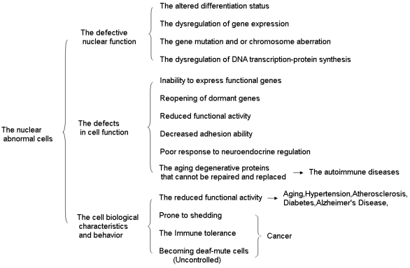
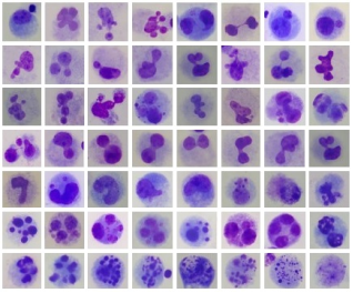
Traditionally, the genetic toxicity damage has generally only referred to the abnormalities of gene structure; while the epigenetics has merely focused on the abnormalities of DNA methylation leading to abnormal gene expression and regulation; both are not comprehensive or complete enough. In fact, the genetic toxicity damage should encompass the entire damage to the cell nucleus, including both chromatin or DNA damage, as well as other molecular damage . Only that the chromatin or DNA damage can lead to gene mutations and/or chromosomal aberrations, mainly affecting the structure of genes; while other molecular damages mainly affect gene expression and regulation (affecting the function of gene switches). Therefore, the genetic toxicity damage refers to damage to the cell nucleus, and conversely, damage to the cell nucleus is just the genetic toxicity damage. The classical genetics only concerned the abnormalities of gene structures, but failed to pay attention to the abnormalities of gene switches, that is, it ignored or overlooked abnormal gene expression and regulation . However, the epigenetics did not notice any abnormalities in the gene structure. Although it observed abnormalities in gene expression regulation (abnormal gene switches), it did only take notice of abnormalities of DNA methylation, and most of the factors that affect gene expression regulation were overlooked . And the genetic mutations, chromosomal aberrations, micronuclei, nuclear abnormalities, and epigenetic changes are all merely the results and manifestations of damage to the cell nucleus or genetic toxicity damage (Figure 1).
Figure 1. Abnormal nuclear cells resulted from the nuclear damage.
4.4. Both Gene Mutations and Chromosomal Aberrations Are Resulted from the Damage of Cellular Nucleus
The chances of radiation and various carcinogenic compounds directly interacting with chromatin or DNA are relatively rare. Instead, they usually first interact with water molecules to generate reactive oxygen and free radicals, which then damage the chromatin or DNA and induce gene mutations and chromosomal aberrations . In addition, the cell nucleus has a powerful free radical clearance system, a DNA damage repair system, and a chromatin structure activity maintenance system. As long as the cell nucleus functions normally, the gene mutations and chromosomal aberrations are generally not likely to occur . Therefore, the gene mutations and chromosomal aberrations are essentially the results and manifestations of damage to the cell nucleus. The chromatin or DNA damage, if it only affects the linear structure of DNA, involves only a few bases, and affects only one gene, is a gene mutation; if it affects the structure and number of chromosomes, involves large segments or multiple fragments or even the entire chromatin, and affects multiple genes, it is a chromosomal aberration . Micro-nuclei are the result and manifestation of chromosomal aberrations (Figure 1).
4.5. There Are Different of Genotoxic Effects in Different Cells
For non-dividing cells such as brain cells, cardiac muscle cells and skeletal muscle cells, since they do not undergo division and proliferation, naturally no daughter cells are produced. Furthermore, since its DNA does not replicate, any damage to its chromatin or DNA will not be passed on to the daughter cells, and thus will not cause genetic toxicity effects; therefore, referring to such non-dividing cells as having genetic toxicity damage might not be appropriate. For cells that can divide, such as hematopoietic stem cells in the bone marrow, dermal cells, intestinal mucosal epithelial cells, and reproductive cells, after DNA replication and cell division, the chromatin or DNA damage will be passed on to the daughter cells, resulting in cytogenetic toxic effects. Among them, damage to the nuclear structure of germ cells can also affect the individual offspring, resulting in genetic toxic effects in organisms . However, the genetic toxicity effects of male and female reproductive cells are different. Since the female eggs remain in the second meiotic division stage during the early stages of the embryo, their chromosomes are in a sealed state for 12 to 100 years. Therefore, the egg DNA is less likely to undergo gene mutations or chromosomal aberrations, and is also less likely to be erased or altered. Male reproductive cells continuously divide to produce sperm, so their DNA is prone to genetic mutations and/or chromosomal aberrations, and is also easily rewritten or altered . This suggests that the experiences and physical change information of human beings in their conquest of nature may have been mainly encoded into the offspring’s genome through male sperm. This implies that the human evolution was probably achieved mainly through males. This does not mean that women are unimportant; rather, they are even more important because the stability of the human genome is mainly achieved through women. This is in line with the principles of evolution and genetics. Women release only one egg each month, and it requires meticulous care and cannot afford any mistakes. While a single sperm is sufficient for fertilization, men produce hundreds of millions of sperm every day. Why do they expend so much energy to produce such a large number of sperm? Because during the process of encoding biological information into sperm DNA, the human body produces a large number of abnormal or defective sperm. Most of these defective sperm are eliminated in the competition of fertilization. Only a sufficient number of sperm can ensure reproductive success and record the information of human physical changes into the genetic code of the offspring, in order to ensure the health of the offspring and promote human evolution. It can be seen that the genetic toxicity effects and outcomes vary depending on the type of cells and the gender of the individuals. When studying or discussing genetic toxicity damage, it is necessary to distinguish whether the cells are dividing, whether they are reproductive cells, and whether they are male or female.
4.6. The Role Gene and Its Corresponding Protein Are the Most Fundamental Biological Markers of Human Body
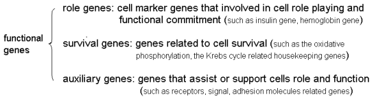
Every cell in the human body contains a complete set of genes, but each cell selectively expresses only a few of these genes while the majority remain inactive. The expressed genes are all closely related to the functions of cells or the human body and are indispensable. They generally play a certain role or undertake a certain function within the body, and thus are called functional genes (dominant genes, FG/DG) . The functional genes mainly include genes related to the survival of the cell itself (survival genes, SG), genes related to the functional role of the cell (role genes, RG), and genes that provide support and assistance (auxiliary genes, AG). The survival genes, such as genes related to enzymes like glycolysis, oxidative phosphorylation, and the tricarboxylic acid cycle, as well as the transcriptional factor genes and so on . The role genes such as hemoglobin and insulin are respectively the role genes of the red blood cells and pancreatic islet cells. The auxiliary genes are rather complex, including carrier channel protein genes, signaling molecule genes, adhesion molecule genes and so on . The survival genes and auxiliary genes are roughly the same or similar in all cells, while the role genes vary greatly in different cells. Therefore, the role genes and their encoding proteins serve as the fundamental markers that distinguish different cells, and they are the primary biological markers for cells or the human body. Other biomarkers are generally derived or generated from that. Insufficient understanding or disregard of this fact may be one of the flaws of the existing biomedical theories (Figures 2, 3).
Figure 2. Genes can be classified into three categories based on their expression states.
Figure 3. Functional genes can be classified into three types based on their functions.
4.7. The Nuclear Damage Results in Cellular Nuclear Abnormalities in the Morphology, Structure and Function
After the damage to the cell nucleus, its morphology, structure and function will all become abnormal; the abnormality of morphology and structure is called nuclear abnormality, and nuclear abnormality can have various forms. A micronucleus is essentially a kind of nuclear abnormality. Traditionally, the nuclear abnormalities are a descriptive or morphological concept, generally not involving nuclear functions; in fact, the nuclear damage can also lead to nuclear functional abnormalities . The abnormalities of nuclear function in dividing cells and non-dividing cells are different. The main functions of the cell nucleus are to replicate, transcribe and synthesize ribosomes, and both transcription and ribosome synthesis aim to produce proteins. Therefore, for non-dividing cells, the nuclear damage mainly causes dysfunction in the process of DNA transcription and protein synthesis. However, for dividing cells, nuclear damage also affects cell division and DNA replication. The synthesis and degradation of proteins are always in a state of dynamic equilibrium. While performing their functions, proteins will continuously undergo denaturation and aging. Therefore, the human body needs to constantly synthesize new proteins for supplementation or replacement . The cellular nuclear damage and dysfunction in DNA transcription-protein synthesis processes prevent the timely replenishment or replacement of degenerated proteins. As a result, various functions of the cells decline; for proliferating cells, it also affects cell division. Eventually, it leads to the decline of cellular and tissue organ functions, human aging, and triggers of various chronic and refractory diseases (Figure 1).
4.8. The Cellular Functional State and Biological Behavior Are Changed by the Nuclear Damage
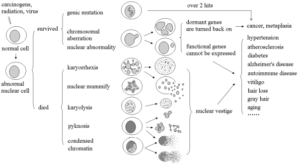
The cellular nuclear damage can affect the expression of functional genes, ultimately influencing the functional state, biological behavior and characteristics of the cells. For instance, defects in the expression of genes of carrier channel receptor and other protein can affect the absorption and transport of cells, as well as the resting potential, osmotic pressure and signal transduction of cells . Disorder in the expression of adhesion molecule genes will affect the cell adhesion connections, leading to cell detachment and migration. Disorder in the expression of signal molecule genes can affect cell communication and interaction, leading to poor responses in neuroendocrine regulation and even resulting in deaf and mute cells that are not under the control of the body . The abnormal nuclear cells (ANC) do not express foreign proteins or antigens, and therefore are not recognized or eliminated by the immune system of the body. Some human genes are continuously expressed, some are intermittently expressed, and some never express at all throughout one's lifetime. The genes that never express at all are called sealed genes, dormant genes or recessive genes. For instance, the hemoglobin gene is a role gene (a functional gene) in red blood cells, but it acts as a sealed gene in brain cells and pancreatic cells. The genes related to cell division and proliferation are not expressed in most cells, remaining in a dormant state (i.e., sealed genes), and are considered as the basic biomarkers of tumors . The cellular nuclear damage may cause the reactivation of sealed genes. If genes related to cell division and proliferation are reactivated, it can lead to the development of cancer or metaplasia. Therefore, the biological behaviors of abnormal nuclear cells mainly manifest as: functional decline, easy detachment, immune tolerance, uncontrolled growth, easy carcinogenesis or metaplasia, and loss of contact inhibition function and so on (Figure 4).
Figure 4. Biological characteristics and behavior of the abnormal nuclear cells.
4.9. The Cellular State of Differentiation Is Disrupted by the Nuclear Damage
All cells in the human body are essentially differentiated cells. The process from the fertilized egg to mature somatic cells is called differentiation. The differentiation is actually the process of modifying cells and altering the gene expressive profile which is the process of silencing the irrelevant genes and opening functional genes. The cell modification, changes in gene expression profiles, and the formation of tissues, organs and embryos are all achieved simultaneously as a whole, among which the change in gene expression profiles serves as the prerequisite and foundation . Which genes are expressed by each cell, how they are expressed, when they are expressed, who regulates them, and how they are regulated - all of these have already been determined or pre-ordained during the process of differentiation. This means that the expression state of genes or the gene expression profile is already determined during differentiation and is irreversible; it also implies that the regulation of gene expression has been simplified and optimized, and being patterned, programmed and automated. In fact, all the various functional activities of human cells are carried out and completed on the basis of differentiation, so cells must maintain a differentiated state . The so-called maintaining the differentiated state means ensuring that functional genes are expressed promptly and that dormant genes remain in a sealed state at all times. The cellular nuclear damage can disrupt the differentiation state, alter the gene expression profile or gene expression status, resulting in the inability of functional genes to be expressed, the activation of dormant genes, and the situation where genes being needed fail to be expressed while genes that should not be expressed are expressed. The failure of functional genes to express will affect the functional state and biological behavior of cells, and the activation of dormant genes can lead to cancer (Figures 4, 5).
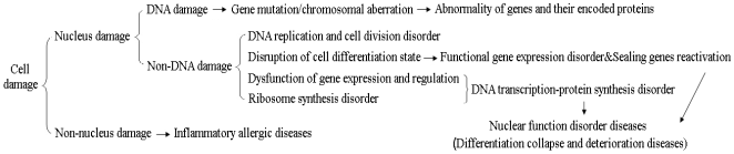
Figure 5. The nuclear damage is different from the non-nuclear damage.
4.10. The Stem Cells Are Difficult to Be Used to Recreate Human Organs
The humans have confused cell division with cell differentiation. Some human cells divide, some do not, some divide along with differentiation, and some only divide without differentiation. For instance, the dermal cells, intestinal mucosal epithelial cells, and mesenchymal stem cells, only divide but do not differentiate. They essentially do not belong to the stem cells (SC) but are part of the life cycle, similar to the single-celled organisms. The embryonic stem cells and bone marrow hematopoietic stem cells both divide and differentiate. Although they possess the characteristics of stem cells, they all differentiate towards specific targets, and require specific molecules for regulation. The humans cannot change their differentiation targets and directions, and it is even more impossible for them to differentiate according to human wishes. The differentiation is actually the process of modifying cells and altering the gene expression profile, which is the process of silencing irrelevant genes and expressing functional genes. After differentiation, the gene expression profile generally remains unchanged, namely maintaining the differentiated state. The disruption of the differentiation state may lead to aging and trigger complex diseases, such as cancer, degenerative diseases, Alzheimer's disease, autoimmune diseases, diabetes, hypertension, atherosclerosis and so on. It is impossible for humans to use stem cells to differentiate and recreate human organs, unless humans master the regulation of gene expression. The so-called stem cell therapy in clinical practice mostly utilizes the paracrine effects of stem cells rather than their differentiation functions . In Japan, mesenchymal stem cells have been also renamed as mesenchymal matrix cells. The so-called stem cells currently used in clinical applications are essentially the abnormal nuclear cells, which is induced under the influence of drugs or chemical reagents by the damage to the cell nucleus, disruption of the differentiation state and changes in the gene expression profile. During DNA replication, it cannot serve as a template for transcription, and during transcription, it cannot act as a template for replication; therefore, replication and transcription are contradictory. Due to the contradiction of replication and transcription, if terminally differentiated cells such as brain cells and cardiac muscle cells divide, it will lead to the disintegration of these tissues and organs into cell homogenates. The human body will disintegrate, and multicellular organisms including us humans will become extinct.
Human body repair includes native repair and alternative repair. The native repair is also known as self-renewal, which includes molecular renewal and cellular renewal. The molecular renewal mainly involves the synthesis of new identical molecules to replace the abnormal ones. For instance, the new proteins are synthesized to replace the aged and denatured identical protein molecules. The terminal differentiated cells such as brain cells, cardiac muscle cells, and skeletal muscle cells mainly undergo repair through molecular renewal. The cell renewal mainly involves replacing aged cells with new cells of the same type, such as blood cells, epidermal cells, and intestinal mucosal epithelial cells. The alternative repair mainly refers to the process of repairing using another type of cell, that is, replacing the original cells with a different kind of cell. The alternative repair in human body mainly involves the use of mesenchymal stem cells (fibroblasts, glial cells, type II lung cells) for repair, namely scar repair; it can be applied to the repair of most tissues and organs, but it probably leads to a decrease in organ function. The organs such as brain, heart, muscles, eyes, lungs and kidneys mainly undergo repair through the molecular renewal. After damage, they are mainly repaired through scar tissue and this affects the functions of the organs. These types of organs cannot be repaired through cell renewal; otherwise, it would damage the structure of the tissues and organs, leading to their disintegration. However, the organs such as the skin, blood cells and intestinal mucosa, do not need to undergo molecular renewal for repair, because these types of cells die shortly after completing their tasks. It is impossible for humans to repair tissues and organs through stem cell differentiation unless humans master the regulation of gene expression. Because the stem cells cannot differentiate as humans expect and desire, and humans currently cannot control the direction and path of stem cell differentiation. The stem cell is probably a false proposition, and the research current in regenerative medicine might be futile.
5. The Nuclear Damage Is Different from the Non-nuclear Damage
The cellular nucleus damage is mainly caused by the radiation, viruses, and various carcinogenic compounds, while the non-nuclear damage (NND) is mainly caused by infections, poisoning, and allergies. The cellular nucleus damage is mainly of chronic type, usually without inflammatory manifestations; the non-cellular nucleus damage is mainly of acute or subacute type, often accompanied by inflammatory allergic reactions. The cellular nucleus damage probably main causes various chronic and refractory diseases, while the non-cellular nucleus damage probably main cause inflammatory and allergic diseases. The traditional cytotoxic damage is mainly non-nuclear damage. The theoretical basis of current biomedical theories is based on the non-nuclear damage, and the diseases discussed are mainly inflammatory and allergic diseases . Although the genetic effects resulting from gene mutations and/or chromosomal aberrations were noted, the mechanism of the genetic toxicity damage was not explored on an overall cellular level. Therefore, the causes and mechanisms of these chronic and refractory diseases have not been comprehensively and systematically understood. The human diseases include nuclear dysfunction disorders mainly caused by damage to the cell nucleus, as well as inflammatory and allergic diseases mainly caused by non-nuclear damage. The factors causing nuclear damage are mainly genotoxic damage. While non-genotoxic factors may also directly or indirectly affect the function of the nuclear, they generally do not produce genotoxic effects . It is not denied that in many cases, the two factors often coexist, interweave with each other, and jointly influence the progression and outcome of the disease. The cell nucleus, endoplasmic reticulum, Golgi apparatus and so on, all participate in protein synthesis. Among them, gene expression and regulation are the core and key steps. Therefore, damage to the cell nucleus is of paramount importance. As long as the function of the cell nucleus is normal, the damage to other cellular organelles can generally be repaired or replenished. The cellular damage is precisely the link between genetics and the environment, representing the interaction between these two factors. The so-called polygenic diseases are not actually caused by multiple genes; rather, they are the result of cell nuclear damage leading to disorders in gene expression regulation (Figures 4, 5).
6. Analysis of the Pathogenicity of the Genotoxicity
A large number of reports suggest that gene mutations and/or chromosomal aberrations are the main causes of cancer . Numerous reports have shown that the incidences of micronuclei, nuclear abnormalities, and DNA methylation abnormalities among cancer patients have significantly increased . There are also reports suggesting that chronic and refractory diseases such as aging, hypertension, atherosclerosis, diabetes, Alzheimer's disease, degenerative diseases, and autoimmune diseases are also related to the aforementioned genetic toxic damage . In fact, a gene represents only a single peptide chain or a protein. Therefore, the gene mutations typically affect only one gene or protein, mainly causing single-gene diseases such as hemophilia and thalassemia . Some genes are expressed while others are not. The genes that are expressed are the functional genes, while the non-expressed genes are mainly the genes sealed (the dormant genes). The gene mutations that are sealed off generally do not cause diseases. Due to the different functions of various genes, the pathogenicity of functional genes and the degree of their pathogenicity also vary. The gene mutations of enzyme protein mainly affect metabolic or biochemical reactions. The structural gene mutations can influence the morphology and compartmentalization of cells, while the role gene mutations can affect the role-playing of cells. The chromosomal aberrations, due to involving a large number of genes and proteins, often cause chromosomal diseases and birth defects, and may also lead to miscarriage and stillbirth . Epigenetic alterations mainly refer to abnormal regulation of gene expression, which may lead to disorders in the expression of a series of genes or alterations in their levels. Among them, the inability of functional genes to express will affect the functional state and biological behavior of cells, causing aging and triggering various chronic and refractory diseases; while dormant genes reactivating can cause cancer (Figures 4-6).
As people age, the number of abnormal nuclear cells increases while the number of normal cells decreases. Most cells of the elderly have transformed into abnormal nuclear cells. As a result, the functions of tissues and organs decline, eventually leading to human aging and the development of various chronic and intractable diseases. These diseases may not be caused by genetic mutations or chromosomal aberrations, but rather by damage to the cell nucleus, disruption of differentiation state, dysfunction of gene expression and regulation, as well as disorders in DNA transcription-protein synthesis processes . Genetic mutations and chromosomal aberrations are merely the consequences and manifestations of nuclear damage, and they are accompanying phenomena of the aforementioned diseases. These diseases all fall under the category of degeneration or deficiencies in the function of the cell nucleus (diseases of nuclear function failure, or collapse of state of differentiation) (Figures 4-6).
Figure 6. The chronic intractable diseases are probably derived from the nuclear damage.
7. The Current Situation
Due to the insufficient understanding of genetic toxicity damage, the existing biomedical theories and research may have deficiencies or flaws. For instance: Confusing single-celled organisms' cells with multi-cellular organisms' cells (differentiated cells), confusing cell survival with cell division and proliferation, and confusing the death of single-celled organisms with the life, aging and death of multi-cellular organisms ; Only chromatin or DNA was observed, but the cell nucleus as a whole was not noticed; only genes were concerned, but gene expression regulation was not paid attention; only abnormal gene structures (abnormal DNA gene sequences) were concerned, but abnormal gene switches were not discussed . Only focuses on genetic abnormalities while neglecting differentiation abnormalities; only concentrates on static and isolated molecules while ignoring dynamic molecular interactions and molecular networks. We only know that differentiation gives rise to tissues, organs and embryos, but we don't know that it also remodels cells, alters genetic profiles of expression, and changes regulatory patterns of gene expression. Only knows those inflammatory allergic diseases characterized by cell toxicity damage (non-nuclear damage), but do not knows the diseases of nuclear dysfunction or disorders induced mainly by nuclear damage . It is not genes that control life; rather, it is the natural structure and molecular properties of nucleic acids that control life and the biological world, including us humans. It is nucleic acids working together with proteins that create life, build the organisms, and shape us humans. Most of the current biomedical research data or knowledge come from cultured cells in vitro. These cells have long transformed into single-cell biological cells (with altered gene expression profiles). Therefore, using such cells to explain human differentiated cells is clearly unscientific and even incorrect. Cancer may not be caused by genetic mutations or chromosomal aberrations. The most popular telomerase theory and cell division limit theory regarding aging might be incorrect, and the theoretical hypotheses regarding other chronic and refractory diseases may also have flaws . There are also significant deficiencies in the clinical treatment of chronic and refractory diseases. What is particularly worrying is that for the treatment of these complex diseases, long-term or even lifelong medication is often required. The fundamental reason is that the majority of drugs do not act on pathological cells (cells with abnormal nuclei), but instead alleviate symptoms by interfering with the functions or metabolism of normal cells, disrupting the homeostasis or balance of the cell or organism. This is actually only a symptomatic treatment, which not only has poor efficacy but may also harm health and shorten lifespan.
8. The Expectation
The current medical system is a "static medicine" that relies on the "gene/molecule-biological body" as its basis, completely ignoring the cell which is the cornerstone and link. It is urgent today to establish a "dynamic medicine" model or concept based on the theoretical foundation of "gene/molecule - cell - organism". The static medicine attempts to directly explain the entire human body and all diseases using individual genes or molecules. This is equivalent to extracting a single gene/molecule, in an attempt to explain the constantly dynamic and interacting networks of cells. Therefore, it is very difficult to provide a reasonable explanation for the aforementioned chronic and refractory diseases. The "dynamic medicine" model explains cells at the molecular level, and then uses cells as the foundation to explain the human body and diseases; it takes nuclear damage and gene expression regulation as the entry points, starting from analyzing the functional state and biological behaviors of abnormal nuclear cells to explain diseases and aging; thus, it can better reveal the causes and mechanisms of the aforementioned complex diseases. A systematic analysis and review of genetic toxicity damage are conducted, aiming to enhance people's understanding and attention, providing theoretical support for the prevention and treatment of occupational injuries and related complex diseases, and also offering new ideas and methods for occupational health research.
Abbreviations

GM

Gene Mutation

CA

Chromosomal Aberration

MN

Micronucleus

NAM

Nuclear Abnormality

GTD

Genotoxic Damage

SM

Static Medicine

DM

Dynamic Medicine

BM

Biological Marker

CD

Chromosomal Damage

NCD

Non-chromosomal Damage

AGST

Abnormal Gene Structure

AGSW

Abnormal Gene Switche

ND

Nuclear Damage

NND

Non-nuclear Damage

ANC

Abnormal Nuclear Cell

FG/DG

Functional Gene / Dominant Gene

SG

Survival Gene

RG

Role Gene

AG/HG

Auxiliary Gene / Helper Gene

SC

Stem Cell

Acknowledgments
I thank my director, the professor Rouli Zhou, for providing my lots of meaningful thoughts.
Author Contributions
Maojin Li: Conceptualization, Investigation, Methodology, Visualization, Writing – original draft, Writing – review & editing
Data Availability Statement
The data that support the opinions of this review are openly available.
Conflicts of Interest
The author declares there is no conflict of interest.
References
[1] Haiyan ZHU, Guangning SU, Zhike TAN, Dishun HUANG. Characteristics of nitrosamine pollution in drinking water of Baiyun Section of Liuxi River [J]. China Preventive Medicine, 2024, 25(08): 1028-1034.
[2] Hairong ZHANG. Effects Assessment of Frying Conditions on Benzopyrene in Edible Oil [J]. China Food Safety Magazine, 2024, (16): 135-138+144.
[3] Xiuting LI, Hongbing ZHANG, Yuhua TANG, Qiu DONG, Shizhi WANG, et al. Analysis of occupational health examination results of radiological workers in a city in 2023 [J]. Chinese Journal of Clinical Research, 2025, 38(06): 955-959.
[4] Huaxian WANG, Chang LIU, Youyou WANG, Huahui BIAN, Bingqing ZENG, et al. Analysis of lens opacification status and influencing factors among radiological workers in China’ s nuclear power plants [J]. Radiation Protection, 2025, 45(03): 266-272.
[5] Ortega Pijeira MS, Magne TM, da Silva NCG, Ramos Ribeiro ERF, Albuquerque Silva YJ, et al. Ionizing Radiation: Chemical Kinetics, Chemical Bounds, and Radiation Chemistry on Polymers [J]. Curr Top Med Chem. 2023; 23(15): 1414-1424.
[6] Ishikawa Y, Ushijima Y, Kiyoi H. Recent advances in AML with mutated NPM1 [J]. Int J Hematol, 2024, 120(5): 556-565.
[7] Al-Jumayli M, Brown SL, Chetty IJ, Extermann M, Movsas B. The Biological Process of Aging and the Impact of Ionizing Radiation [J]. Semin Radiat Oncol, 2022, 32(2): 172-178.
[8] Aragane Y, Higashino T, Kinoshita K, Ashenagar MS, Higashino H. Hypertension-Associated Genes in the Mesenteric Artery of Three Spontaneously Hypertensive Rat Substrains Identified Using a DNA Array Method [J]. Front Biosci (Landmark Ed), 2022, 27(6): 191.
[9] Olga V. Kryukova, Igor O. Islanov, Elena V. Zaklyazminskaya, et al. Effects of Angiotensin-I-Converting Enzyme (ACE) Mutations Associated with Alzheimer’s Disease on Blood ACE Phenotype [J]. Biomedicines, 2024, 12(10): 2410-2410.
[10] Tyagi N, Uppili B, Sharma P, Parveen S, Saifi S, et al. Investigation of RFC1 tandem nucleotide repeat locus in diverse neurodegenerative outcomes in an Indian cohort [J]. Neurogenetics, 2024, 25(1): 13-25.
[11] Baosheng YANG, Gang LI, Haiyan JIAO, Li LI, Lan LUO, et al. Medical Genetics [M], 2rd ed.; Beijing Higher Education Press, Beijing, China. 2019, PP: 12-19.
[12] De Wolf D, Singh K, Chuah MK, VandenDriessche T. Hemophilia Gene Therapy: The End of the Beginning? [J] Hum Gene Ther, 2023, 34(17-18): 782-792.
[13] Huixin YANG, Shuyan WEI, Xiaoyan WEN. Analysis of the prenatal diagnosis indications and pregnancy outcomes of 76 fetuses with sex chromosome aneuploidy [J]. Chinese Journal of Family Planning, 2024, 32(07): 1698-1702+1706.
[14] Ma Y, Guo L, Fang L, Hou D, Chen R, et al. Assessment of radiation doses and DNA damage in pediatric patients undergoing interventional procedures for vascular anomalies [J]. Mutat Res Genet Toxicol Environ Mutagen. 2023, 889: 503653.
[15] Nan MA, Junchao FENG, Yulong LIU, Jie LI, Yu GAO. Analysis of radiosensitivity indicators for radiation workers at a nuclear power plant [J]. Chinese Journal of Radiological Health, 2025, 34(02): 264-269.
[16] Maojin LI, Anyi HU. The research progress and biological significance of abnormality of cell nucleus [J]. China Occupational Medicine, 2021, 48(6): 708-711.
[17] Janssen AFJ, Breusegem SY, Larrieu D. Current Methods and Pipelines for Image-Based Quantitation of Nuclear Shape and Nuclear Envelope Abnormalities [J]. Cells. 2022, 11(3): 347.
[18] Smith ZD, Hetzel S, Meissner A. DNA methylation in mammalian development and disease [J]. Nat Rev Genet. 2025, 26(1): 7-30.
[19] Bhootra S, Jill N, Shanmugam G, Rakshit S, Sarkar K. DNA methylation and cancer: transcriptional regulation, prognostic, and therapeutic perspective [J]. Med Oncol. 2023 Jan 5; 40(2): 71.
[20] Datta RR, Akdogan D, Tezcan EB, Onal P. Versatile roles of disordered transcription factor effector domains in transcriptional regulation [J]. FEBS J. 2025, 292(12): 3014-3033.
[21] Fendler NL, Ly J, Welp L, Lu D, Schulte F, et al. Identification and characterization of a human MORC2 DNA binding region that is required for gene silencing [J]. Nucleic Acids Res. 2025, 53(4): gkae1273.
[22] Gong P, Guo Z, Wang S, Gao S, Cao Q. Histone Phosphorylation in DNA Damage Response [J]. Int J Mol Sci. 2025, 26(6): 2405.
[23] Tyagi W, Das S. Temporal regulation of acetylation status determines PARP1 role in DNA damage response and metabolic homeostasis [J]. Sci Adv. 2024, 10(42): eado7720.
[24] Barbosa F Jr, Rocha BA, Souza MCO, Bocato MZ, Azevedo LF, et al. Polycyclic aromatic hydrocarbons (PAHs): Updated aspects of their determination, kinetics in the human body, and toxicity [J]. J Toxicol Environ Health B Crit Rev. 2023, 26(1): 28-65.
[25] Suyi WANG, Li SONG, Zhifeng LIU, Rongyue JIANG, Yue SONG, et al. Research progress on epigenetic changes induced by ionizing radiation [J]. Journal of Preventive Medicine, 2025, 37(04): 361-364.
[26] Meinan YAO, Runqiu YU, Weiwei JIANG, Xiaozhe TANG, Bin BAI, Yongzhong MA. Current status, influencing factors and management challenges of indoor radon levels [J]. Occupation and Health, 2025, 41(13): 1867-1872.
[27] Suya ZHENG, Ye CHEN, Zhipeng ZHU, Nan LI, Chunyu HE, et al. Exploiting targeted degradation of cyclins and cyclin-dependent kinases for cancer therapeutics: a review [J]. Journal of Zhejiang University-SCIENCE B (Biomedicine & Biotechnology), 2025, 26(8): 713-739.
[28] Yan YAN, Yi ZHANG. Mechanism of action of the RNA-binding protein CELF1 in diseases [J]. Journal of China Medical University, 2025, 54(08): 673-677.
[29] Huimin QIU, Hengwen ZHANG, Shuyu YUAN, Kejing ZHANG, Jiahui DING, et al. Shenmai San exerts protective effects on the hematopoietic system of irradiated mice by inhibiting apoptosis through the PI3K/AKT pathway [J]. Lishizhen Medicine and Materia Medica Research, 2025, 36(02): 258-265.
[30] Jiabin HU, Youliang ZHOU. Prognostic value and regulatory analysis of nucleoporin Nup93 complex genes in hepatocellular carcinoma [J]. Zhejiang Journal of Integrated Traditional Chinese and Western Medicine, 2025, 35(08): 712-719.
[31] Issa JP. The Two-Hit Hypothesis Meets Epigenetics [J]. Cancer Res. 2022, 82(7): 1167-1169.
[32] de Lima JD, de Paula AGP, Yuasa BS, de Souza Smanioto CC, da Cruz Silva MC, et al. Genetic and Epigenetic Regulation of the Innate Immune Response to Gout. Immunol Invest [J]. 2023, 52(3): 364-397.
[33] Sinkala M. Mutational landscape of cancer-driver genes across human cancers [J]. Sci Rep. 2023, 13(1): 12742.
[34] Kataoka K, Saito Y. Multiple Mutations within Individual Oncogenes: Examples and Clinical Implications [J]. Keio J Med. 2023, 72(3): 88-92.
[35] Skinner MK. Epigenetic biomarkers for disease susceptibility and preventative medicine [J]. Cell Metab, 2024, 36(2): 263-277.
[36] Han TS, Kim DS, Son MY, Cho HS. SMYD family in cancer: epigenetic regulation and molecular mechanisms of cancer proliferation, metastasis, and drug resistance [J]. Exp Mol Med, 2024, 56(11): 2325-2336.
[37] Ziyi DING, Zhao XU, Jingwen CAO, Taotao JI, Yuanyuan ZHOU, et al. Low dose tritiated water induced radiation-adaptive responses in lung cancer cells based on the Nrf2 pathway [J]. Radiation Protection, 2025, 45(01): 67-76.
[38] Liangming REN. Study on amelioration of irradiation-induced salivary gland damage in mice by antioxidants and underlying mechanisms [D]. Peking Union Medical College, 2023.
[39] Xing LI, Ming LIU, Haihui LIN, Yinghong WEI, Shibiao SU. Research progress on the effects of low dose radiation on DNA damage repair [J]. China Occupational Medicine, 2024, 51(06): 700-706+712.
[40] Chen C, Luo Y, Hou X, Li T. Clinical characterization of epilepsy in children with chromosomal aberration 47, XXY [J]. Brain Behav, 2023, 13(8): e3178.
[41] Ay C, Frenzel L, Pinachyan K, Le Quellec S. Gene therapy for haemophilia A and B, from basic principles to clinical implementation: An illustrated review [J]. Haemophilia, 2024, 30(1): 5-15.
[42] Murthy A, Tirado CA. Fanconi Anemia, AML, and MDS [J]. J Assoc Genet Technol. 2024; 50(2): 64-68. PMID: 38824651.
[43] Dashti P, Lewallen EA, Gordon JAR, Montecino MA, van Leeuwen JPTM, et al. Protein arginine methyltransferases PRMT1, PRMT4/CARM1 and PRMT5 have distinct functions in control of osteoblast differentiation [J]. Bone Rep. 2023, 19: 101704.
[44] Choubey P, Kaur H, Bansal K. Modulation of DNA/RNA Methylation Signaling Mediating Metabolic Homeostasis in Cancer [J]. Subcell Biochem. 2022, 100: 201-237.
[45] Zhang QQ, Qu Y. Brain-derived neurotrophic factor in degenerative retinal diseases: Update and novel perspective [J]. J Neurosci Res. 2023, 101(10): 1624-1632.
[46] Shufan CAI. Research on Methods for Predicting Ionizing Radiation Exposure Risk Based on Blood Molecules [D]. Lanzhou University, 2025.
[47] Xingyu LIU, Tao WANG, Changjing ZUO, Jianming CAI. Application of non-coding RNA and proteomics in radiation biodosimetry [J]. Academic Journal of Naval Medical University, 2024, 45(12): 1540-1545.
[48] Manda NK, Golla U, Sesham K, Desai P, Joshi S, et al. Tuning between Nuclear Organization and Functionality in Health and Disease [J]. Cells. 2023, 12(5): 706.
[49] Morimoto RI. Cell-Nonautonomous Regulation of Proteostasis in Aging and Disease [J]. Cold Spring Harb Perspect Biol. 2020, 12(4): a034074.
[50] Chaudhary MR, Chaudhary S, Sharma Y, Singh TA, Mishra AK, et al. Aging, oxidative stress and degenerative diseases: mechanisms, complications and emerging therapeutic strategies [J]. Biogerontology, 2023, 24(5): 609-662.
[51] Leńska-Mieciek M, Madetko-Alster N, Alster P, Królicki L, Fiszer U, Koziorowski D. Inflammation in multiple system atrophy [J]. Front Immunol. 2023, 14: 1214677.
[52] Yun SHA, Shihao SHAO, Jinyuan YANG, Beining TANG, Yuhang QUAN, et al. Elucidation of the molecular mechanisms of TRPA1 channel protein in pain modulation and advancements in the development of antagonists for cancer pain management [J]. Chinese Journal of Oncology Prevention and Treatment, 2025, 17(04): 516-522.
[53] Lin WY, Dong YL, Lin Y, Sunchuri D, Guo ZL. Potential role of G protein?coupled receptor 124 in cardiovascular and cerebrovascular disease (Review) [J]. Exp Ther Med. 2024, 29(1): 2.
[54] Wenjuan ZHANG, Tingting LIU, Xiaoli LV, Mengdi ZHANG, Yuxia HU, et al. Study on the Antagonistic Effect of Quercetin on the Action of Benzo [a] pyrene on the AhR-CYP Signaling Pathway [J]. Journal of Inner Mongolia Medical University, 2025, 47(03): 246-250.
[55] Lifang REN, Tuya HASI. Analysis of the changes and diagnostic value of serum D-dimer, alpha-fetoprotein, carcinoembryonic antigen and carbohydrate antigen 199 levels in patients with epithelial ovarian cancer [J]. Chinese Journal of Human Sexuality, 2025, 34(08): 73-76.
[56] Bin WU. The promoting effect of cancer-associated fibroblasts after ionizing radiation on the malignant biological behavior of lung cancer cells and its mechanism [D]. Wuhan University, 2021.
[57] Jackson BT, Finley LWS. Metabolic regulation of the hallmarks of stem cell biology [J]. Cell Stem Cell. 2024, 31(2): 161-180.
[58] Zahoor A, Khazer R, Mehraj I, Gani U, Fayaz F, et al. Aberrant DNA methylation as a key modulator of cell death pathways: insights into cancer progression and other diseases [J]. Funct Integr Genomics. 2025, 25(1): 50.
[59] Xinghua ZHOU. The effect and mechanism of exosomes derived from human amniotic fluid stem cells on hair regeneration after ionizing radiation [D]. Soochow University, 2021.
[60] Thorp EB, Filipp M. Contributions of Inflammation to Cardiometabolic Heart Failure with Preserved Ejection Fraction [J]. Annu Rev Pathol. 2025, 20(1): 143-167.
[61] Okano M, Yamada M, Oka A. Personalized Medicine in Chronic Rhinosinusitis: Treatable Traits Using Biologics for Unmet Needs [J]. Allergy Asthma Immunol Res. 2025, 17(1): 8-21.
[62] Zhu J, Meganathan I, MacAruthur R, Kassiri Z. Inflammation in Abdominal Aortic Aneurysm: Cause or Comorbidity? [J] Can J Cardiol. 2024, 40(12): 2378-2391.
[63] Ogger PP, Murray PJ. Dissecting inflammation in the immunemetabolomic era [J]. Cell Mol Life Sci. 2025, 82(1): 182.
[64] Tornesello ML. TP53 mutations in cancer: Molecular features and therapeutic opportunities (Review) [J]. Int J Mol Med. 2025, 55(1): 7.
[65] Lee AV, Nestler KA, Chiappinelli KB. Therapeutic targeting of DNA methylation alterations in cancer [J]. Pharmacol Ther. 2024, 258: 108640.
[66] Huang D, Shang W, Xu M, Wan Q, Zhang J, et al. Genome-Wide Methylation Analysis Reveals a KCNK3-Prominent Causal Cascade on Hypertension [J]. Circ Res. 2024, 135(3): e76-e93.
[67] Lizama BN, Kahle J, Catalano SM, Caggiano AO, Grundman M, Hamby ME. Sigma-2 Receptors-From Basic Biology to Therapeutic Target: A Focus on Age-Related Degenerative Diseases [J]. Int J Mol Sci. 2023, 24(7): 6251.
[68] Pisetsky DS. Pathogenesis of autoimmune disease [J]. Nat Rev Nephrol. 2023, 19(8): 509-524.
[69] Abed S, Ebrahimi A, Fattahi F, Kouchakali G, Shekari-Khaniani M, Mansoori-Derakhshan S. The Role of Non-Coding RNAs in Mitochondrial Dysfunction of Alzheimer's Disease [J]. J Mol Neurosci. 2024, 74(4): 100.
[70] Elendu C, Amaechi DC, Alakwe-Ojimba CE, Elendu TC, Elendu RC, et al. Understanding Sickle cell disease: Causes, symptoms, and treatment options [J]. Medicine (Baltimore), 2023, 102(38): e35237.
[71] Samelson-Jones BJ, Small JC, George LA. Roctavian gene therapy for hemophilia A [J]. Blood Adv, 2024, 8(19): 5179-5189.
[72] Butler G, Srirangalingam U, Faithfull J, Sangster P, Senniappan S, Mitchell R. Klinefelter syndrome: going beyond the diagnosis [J]. Arch Dis Child, 2023, 108(3): 166-171.
[73] Hokland P, Daar S, Khair W, Sheth S, Taher AT, et al. Thalassaemia-A global view [J]. Br J Haematol, 2023, 201(2): 199-214.
[74] Nie M, Oravcová M, Jami-Alahmadi Y, Wohlschlegel JA, Lazzerini-Denchi E, Boddy MN. FAM111A induces nuclear dysfunction in disease and viral restriction [J]. EMBO Rep. 2021, 22(2): e50803.
[75] Gray PE, David C. Inborn Errors of Immunity and Autoimmune Disease [J]. J Allergy Clin Immunol Pract. 2023, 11(6): 1602-1622.
[76] Gholami A. Alzheimer's disease: The role of proteins in formation, mechanisms, and new therapeutic approaches [J]. Neurosci Lett, 2023, 817: 137532.
[77] Haoyu JI, Hao CHEN, Liang SHI, Meng ZHANG, Ting CHEN, et al. Advances in the Studies of MicroRNA-1976 in Aberrant Cell Cycle Diseases [J]. Acta Academiae Medicinae Sinicae, 2025, 47(04): 644-650.
[78] Xiaofeng NIU, Siyu YANG, Lijuan DENG, Zheng REN, Chengliu BI, Jun YANG. Research progress of cell senescence in the occurrence and development of radiation injury [J]. Radiation Protection Bulletin, 2024, 44(06): 1-9.
[79] Fangjie ZHOU, Longxiang YIN, Ke WANG, Siyuan LUO, Xiaodong WANG. Simulation studies on the characterization of DNA molecular damage induced by protons [J]. Nuclear Electronics & Detection Technology, 2025, 45(06): 804-813.
[80] Zhengyue CAO, Youfeng ZHANG, Zhichun LV, Huiying GAO, Shensi XIANG, et al. CRISPR/Cas9-based knockout library screening identifies BAG3 as a potential regulator of radiosensitivity [J]. Military Medical Sciences, 2025, 49(06): 421-429.
[81] Delemarre T, Bachert C. Neutrophilic inflammation in chronic rhinosinusitis [J]. Curr Opin Allergy Clin Immunol. 2023, 23(1): 14-21.
[82] Goswami D, Anuradha U, Angati A, Kumari N, Singh RK. Pharmacological and Pathological Relevance of S100 Proteins in Neurological Disorders [J]. CNS Neurol Disord Drug Targets. 2023, 22(10): 1403-1416.
[83] Bhatt S, Argueta DA, Gupta K, Kundu S. Red Blood Cells as Therapeutic Target to Treat Sickle Cell Disease [J]. Antioxid Redox Signal, 2024, 40(16-18): 1025-1049.
[84] Lu YR, Tian X, Sinclair DA. The Information Theory of Aging. Nat Aging. 2023, 3(12): 1486-1499.
Cite This Article
  • APA Style

    Li, M. (2026). New Insights into the Mechanisms of Genetic Toxicity Damage. European Journal of Preventive Medicine, 14(3), 45-58. https://doi.org/10.11648/j.ejpm.20261403.12

    Copy | Download

    ACS Style

    Li, M. New Insights into the Mechanisms of Genetic Toxicity Damage. Eur. J. Prev. Med. 2026, 14(3), 45-58. doi: 10.11648/j.ejpm.20261403.12

    Copy | Download

    AMA Style

    Li M. New Insights into the Mechanisms of Genetic Toxicity Damage. Eur J Prev Med. 2026;14(3):45-58. doi: 10.11648/j.ejpm.20261403.12

    Copy | Download

  • @article{10.11648/j.ejpm.20261403.12,
      author = {Maojin Li},
      title = {New Insights into the Mechanisms of Genetic Toxicity Damage},
      journal = {European Journal of Preventive Medicine},
      volume = {14},
      number = {3},
      pages = {45-58},
      doi = {10.11648/j.ejpm.20261403.12},
      url = {https://doi.org/10.11648/j.ejpm.20261403.12},
      eprint = {https://article.sciencepublishinggroup.com/pdf/10.11648.j.ejpm.20261403.12},
      abstract = {The damage of genetic toxicity generally refers to the damage to human genetic material, chromatin or DNA. The genotoxic damage biomarkers such as the gene mutations (GM), chromosomal aberrations (CA), micronucleus (MN), nuclear abnormalities (NAM), and the abnormal DNA methylation are also the results and manifestations of the genotoxic damage (GTD). The genotoxic damage is essentially the cell nuclear damage, which includes not only the chromatin or DNA damage but also the damage to other molecules. The mild DNA damage involving only a few bases and affecting only one gene is called gene mutation. The severe DNA damage involving large fragments or entire chromosomes and affecting multiple genes is called chromosomal aberration. The chromatin or DNA damage mainly affects the gene structure, while other molecular damage mainly affects the gene switching (gene expression and regulation). Traditionally, the genotoxic damage only refers to the abnormal gene structure, while the epigenetics only refers to the abnormality of gene expression and regulation caused by the abnormal DNA methylation. Therefore, both classical genetics and epigenetics are not comprehensive and perfect enough. The nuclear damage (ND) affects not only morphology and structure of cell nucleus but also the function. The nuclear abnormalities usually refer to the abnormalities in the morphology and structure of nuclei. The micronucleus is both a chromosomal fragment and a nuclear abnormality. The abnormal nuclear functions mainly include the abnormalities in replication and transcription, disruption of differentiation status, alteration of profiles of gene expression, and the dysfunction of DNA transcription-protein synthesis. The nuclear damage can affect the functional state and biological behavior of cells, leading to reduced cell function, easy shedding, uncontrollability, immune tolerance, and susceptibility to carcinogenesis or metaplasia. Aging, cancer, hypertension, diabetes, Alzheimer's disease, degenerative diseases, autoimmune diseases and so on, are probably caused by the nuclear damage, and belong to the diseases of nuclear dysfunction. The current biomedicine originates from the cytotoxic damage (essentially non-nuclear damage), and the diseases discussed are inflammatory allergic diseases. Since the genotoxic damage has not been grasped from the perspective of the entire cell nucleus, it is impossible to provide a reasonable explanation for the above-mentioned chronic and refractory diseases. The current biomedicine belongs to "the static medicine (SM)", whose theoretical basis is "the molecular-organism", ignoring the cell as a cornerstone or link. It is necessary to establish a "dynamic medicine (DM)" model or concept based on "the molecular-cell-organism" as the theoretical foundation.},
     year = {2026}
    }
    

    Copy | Download

  • TY  - JOUR
    T1  - New Insights into the Mechanisms of Genetic Toxicity Damage
    AU  - Maojin Li
    Y1  - 2026/07/08
    PY  - 2026
    N1  - https://doi.org/10.11648/j.ejpm.20261403.12
    DO  - 10.11648/j.ejpm.20261403.12
    T2  - European Journal of Preventive Medicine
    JF  - European Journal of Preventive Medicine
    JO  - European Journal of Preventive Medicine
    SP  - 45
    EP  - 58
    PB  - Science Publishing Group
    SN  - 2330-8230
    UR  - https://doi.org/10.11648/j.ejpm.20261403.12
    AB  - The damage of genetic toxicity generally refers to the damage to human genetic material, chromatin or DNA. The genotoxic damage biomarkers such as the gene mutations (GM), chromosomal aberrations (CA), micronucleus (MN), nuclear abnormalities (NAM), and the abnormal DNA methylation are also the results and manifestations of the genotoxic damage (GTD). The genotoxic damage is essentially the cell nuclear damage, which includes not only the chromatin or DNA damage but also the damage to other molecules. The mild DNA damage involving only a few bases and affecting only one gene is called gene mutation. The severe DNA damage involving large fragments or entire chromosomes and affecting multiple genes is called chromosomal aberration. The chromatin or DNA damage mainly affects the gene structure, while other molecular damage mainly affects the gene switching (gene expression and regulation). Traditionally, the genotoxic damage only refers to the abnormal gene structure, while the epigenetics only refers to the abnormality of gene expression and regulation caused by the abnormal DNA methylation. Therefore, both classical genetics and epigenetics are not comprehensive and perfect enough. The nuclear damage (ND) affects not only morphology and structure of cell nucleus but also the function. The nuclear abnormalities usually refer to the abnormalities in the morphology and structure of nuclei. The micronucleus is both a chromosomal fragment and a nuclear abnormality. The abnormal nuclear functions mainly include the abnormalities in replication and transcription, disruption of differentiation status, alteration of profiles of gene expression, and the dysfunction of DNA transcription-protein synthesis. The nuclear damage can affect the functional state and biological behavior of cells, leading to reduced cell function, easy shedding, uncontrollability, immune tolerance, and susceptibility to carcinogenesis or metaplasia. Aging, cancer, hypertension, diabetes, Alzheimer's disease, degenerative diseases, autoimmune diseases and so on, are probably caused by the nuclear damage, and belong to the diseases of nuclear dysfunction. The current biomedicine originates from the cytotoxic damage (essentially non-nuclear damage), and the diseases discussed are inflammatory allergic diseases. Since the genotoxic damage has not been grasped from the perspective of the entire cell nucleus, it is impossible to provide a reasonable explanation for the above-mentioned chronic and refractory diseases. The current biomedicine belongs to "the static medicine (SM)", whose theoretical basis is "the molecular-organism", ignoring the cell as a cornerstone or link. It is necessary to establish a "dynamic medicine (DM)" model or concept based on "the molecular-cell-organism" as the theoretical foundation.
    VL  - 14
    IS  - 3
    ER  - 

    Copy | Download

Author Information
  • Department of Health Examination, Beijing Prevention and Treatment Hospital of Occupational Disease / Beijing Institute of Occupational Disease Prevention and Treatment, Beijing, China
  • Abstract
  • Keywords
  • Document Sections

    1. 1. Introduction
    2. 2. The Common Biomarkers of Genotoxic Damage
    3. 3. The Causes of Genotoxic Damage
    4. 4. Analysis of the Mechanisms of the Genotoxic Damage
    5. 5. The Nuclear Damage Is Different from the Non-nuclear Damage
    6. 6. Analysis of the Pathogenicity of the Genotoxicity
    7. 7. The Current Situation
    8. 8. The Expectation
    Show Full Outline
  • Abbreviations
  • Acknowledgments
  • Author Contributions
  • Data Availability Statement
  • Conflicts of Interest
  • References
  • Cite This Article
  • Author Information