Case Report | | Peer-Reviewed

Acute Puerperal Uterine Inversion: A Rare Obstetric Emergency - Case Studies

Received: 16 February 2026     Accepted: 25 February 2026     Published: 22 July 2026
Views:       Downloads:
Abstract

Introduction: Acute puerperal uterine inversion is a rare yet critical obstetric emergency, commonly during the third stage of labor. The incidence stands at 1 in 20,000 deliveries. Though uncommon poses a serious threat to maternal and fetal wellbeing. The exact etiology remains unclear, factors such as high parity, ligament laxity, uterine abnormalities, abnormal placentation, umbilical cord traction, use of tocolytics, and fundal pressure have been reported as potential risk factors. Not to mention spontaneous cases of puerperal uterine inversion has also been documented. Diagnosis is primarily clinical, though it can be challenging for inexperienced providers, particularly in the case of first or second-degree inversion. Imaging has been used to aid in its diagnosis when in dilemma. Management of acute puerperal uterine inversion aims at restoring and maintaining hemodynamic stability, timely uterus repositioning either manually or surgical. We report two cases of acute puerperal complete uterine inversion, one managed surgically both patients had uneventful recoveries. Conclusion: Acute puerperal uterine inversion is uncommon but a life-threatening condition. Health care provider in delivery rooms should be well trained to promptly diagnose and manage this condition to ensure optimal patient care.

Published in European Journal of Preventive Medicine (Volume 14, Issue 4)
DOI 10.11648/j.ejpm.20261404.12
Page(s) 70-73
Creative Commons

This is an Open Access article, distributed under the terms of the Creative Commons Attribution 4.0 International License (http://creativecommons.org/licenses/by/4.0/), which permits unrestricted use, distribution and reproduction in any medium or format, provided the original work is properly cited.

Copyright

Copyright © The Author(s), 2026. Published by Science Publishing Group

Keywords

Puerperal Uterine Inversion, Obstetrics Emergency, Postpartum Hemorrhage

1. Introduction
Uterine inversion (UI) is a rare catastrophic obstetric entity in need of expedite interventions . It can either be puerperal or non-puerperal type; the former often occurs during the 3rd stage labor . Prevalence of puerperal uterine inversion is reported to be 1 out of 20,000 deliveries . It occurs when the uterine fundus collapse into endometrial cavity, resulting in partially or complete inversion of the uterus . Although the exact cause of uterine inversion is not fully understood, spontaneous occurrence has been reported . Physiological change in pregnancy which are marked by laxity of ligament, hypertrophy of fundus and simultaneous thinning of the lower segment has been postulated as contributing to the pathophysiology of spontaneous occurrence . Never the less primiparous, active pulling of umbilical cord during third stage of labour, short umbilical cord, uterine anomaly has been among reported risk factors .
Occurrence of acute uterine inversion been evidenced across all trimesters and following either vaginal or cesarean birth while the latter being most frequent . The diagnosis UI is a clinical diagnosis though pose a challenge especially if it is first and second-degrees inversion . Imaging modalities such as ultrasound or magnetic resonance imaging (MRI) have been used to aid its diagnosis when time permit or in a clinical dilemma .
Management of acute uterus inversion go hand in hand with prevention and management of major obstetric hemorrhage . Adequate fluid resuscitation, transfusion of blood and blood products are key in correction and maintaining hemodynamic stability . Additionally, tocolytics agents such as magnesium sulphate, beta agonist, halogenated compound has been used in aiding uterus reposition . Non-surgical maneuver such as Johnson’s and O’Sullivan have been applied as first line in most of cases . Other surgical options such as Spinelli, Huntington, Haultain or laparoscopic reposition have being employed to aid it reposition successful . The choice of surgical / surgical approach is individualized depending on patient condition, attending experience and necessary equipment .
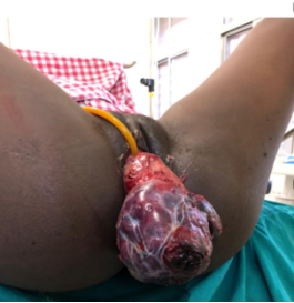
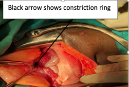
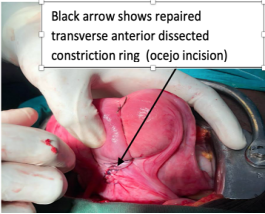
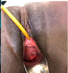
Case 1: An 18-year-old woman, a P0+1, was referred to our hospital with excessive vaginal bleeding and a vaginally protruding mass following a home delivery at 20 weeks of gestation. She had not yet booked for antenatal care. She was diagnosed to have uterine prolapse and initial managements including oxytocin 10IU intramuscular and misoprostol 800mcg per rectum, catheterized, intravenous crystalloids fluids and 2 units of whole blood. Fourteen (14) hours later she was referral for specialized care at our facility. On arrival she was severely pale, tachycardic, she had cold extremities and hypotensive. Blood pressure was 80/42 mmHg and pulse rate was 124 beats per minute. On abdominal examination she had a soft abdomen and uterus was not palpable. On vaginal examination; she had visible inverted uterus with partial placental tissue in situ, there was no active vaginal bleeding at that time as seen in Figure 1. Other systemic examinations findings were essentially normal. She was diagnosed with acute uterine inversion with severe anaemia and hypovolemic shock. A complete blood count revealed leukocytosis of 41.29×10⁹/L, low hemoglobin levels of 4.3 g/dL, hematocrit of 14.3%, and platelets of 159 x 109/L. she is blood group O positive. Other liver, renal, electrolyte and bleeding indices were in normal range. She was resuscitated with intravenous fluids, plasma volume expander a decision was made to proceed with examination under anesthesia. Under general anesthesia induction was done with propofol maintained with halothane. Manual reposition of the inverted uterus was performed using Johnson’s maneuver while portion of placental tissue was in situ was unsuccessful. Decision for abdominal reposition was reached whereby tight constriction ring was identified with complete uterine inversion as shown in Figure 2 below. Successful reposition was achieved by aid of Ocejo incision on constriction ring followed by Hayman’s compression suture as shown in Figure 3. Abdominal closure was done in layer followed by vaginal examination where normal positioned cervix was observed as shown in Figure 4. The patient received 4 unit of blood, intravenous antibiotics, sitz bath with potassium permanganate and analgesics on course of her admission. She had an unremarkable recovery and was discharged home on the tenth day following her hospitalization. She never returned for further evaluation after discharged.
Figure 1. Shows in uterine inversion with partial placental tissue in-situ.
Figure 2. Shows in uterine inversion with con-striction ring and right and left adnexa.
Figure 3. Reposition uterus with Hay-man’s compression suture.
Figure 4. Appearance of the cervix after uterus reposition.
Case 2: A18-year-old she was admitted as P0+1 as self-referral from home. She presented with a complaint of profuse vaginal bleeding accompanied with mass protrusion vaginally for four (4) hours. She had not yet book for antenatal care; on examination she was estimated to be at 24 week’s gestation age. On general evaluation she was afebrile, severely pale, tachycardic and she had cold extremities. Her vital signs; blood pressure of 76/44, mean arterial pressure (MAP) of 57, pulse rate of 116 and oxygen saturation was 89% in room air. On abdominal examination; she had soft, non-tender abdomen and uterus was not palpable abdominally. Digital exam; placenta tissue was in-situ with visible inverted uterine fundus. Other systemic examinations were essentially normal. She was diagnosed with hypovolemic shock secondary to complete acute puerperal uterine inversion. A complete blood count revealed normal WBC of 10.5×10⁹/L, low hemoglobin levels of 8.2 g/dL, hematocrit of 25.3%, and platelets of 189×109/L, B-human chorionic gonadotrophic hormone (B-HCG)was more than 50, 000. Electrolyte; (sodium, potassium and chloride) were within the normal ranges. Bed side clotting time was 6 minutes. She was resuscitated with crystalloids fluids, plasma volume expander, and oxygen 8L via face mask. Oxytocin 10IU intramuscular was given and she was catheterized. Examination under general anesthesia was done with propofol as induction agent maintained with halothane. Uterus was manually repositioned using Johnsons maneuver while the placental tissue was in-situ followed by manual removal of placental tissue and blunt curettage to ensure complete removal of placental tissue. Four (4) foley balloon catheters were inserted and ballooned with 30mls to 40mls of normal saline each as uterine tamponade. Misoprostol 800 mcg was inserted per rectally and oxytocin 20IU in 500mls of normal saline was titrated for four (4) hours. The estimated blood loss was around 800mls. Twelve (12) hours post uterus reposition the foley catheter were removed and there was no active vaginal bleeding. Uterus was well contracted on abdominal examination. She received 1 unit of whole blood, intravenous antibiotics, sitz bath with potassium permanganate and analgesics on course of her treatment. On seventh day following admission, she was discharge home with unremarkable recovery.
2. Discussion
Acute puerperal uterine inversion is quite uncommon with incidence reported to be 1 out of 20,000 deliveries . Though rare but it is one of the fatal obstetrics emergencies and caries potential threat to life of the mother and the fetus . In our setting we attended (2) two cases in duration of one month apart with acute uterine inversion. In both case patients presented in hypovolemic shock and mass protrusion vaginaly following home delivery. Similar presentation was reported in other cases . The diagnosis of puerperal acute uterine inversion can be challenging especially in unexperienced clinician . In case 1, there was misdiagnosis of intrapartum uterine prolapse which necessitated referral for more than 100km for obstetrician review in shock she merely survived. Clinical evaluation remains a main stay for its diagnosis; however, in clinical dilemma or when time permit imaging can be considered . In our cases the imaging was not need as clinical diagnosis was certain.
Timely diagnosis and management are keys to reduce associated morbidity and mortality . The goal is to restore and maintain hemodynamic stability followed either manual reposition or surgical reposition by laparotomy or laparoscopy . Immediately reposition has been reported to be more successful due to less edema resulting into incarceration of the uterus . This was observed in case 2 contrary to case 1. The success rate of manual reposition is reported ranging from 43%-88% in ideal situation . Among the evidenced risks for failure are such as tight constriction ring, placental morbidity anomaly, inadequate skills , where the former was observed in case 1. Failure of manual revision was also reported in other cases . The other attractive manual reposition technique is hydrostatic reduction (O’Sullivan), but in our case 1 we did not opt for that due to tight constriction ring and edematous uterus which carries higher chance of failure.
Surgical reposition has been reported successful without making uterine incision (Huntington maneuver) . However, the maneuver carries risk of round ligament tear . In our case 2, we did not perform huntington maneuver due to tight constriction ring and fear of round ligament injuries. Transverse incision was made along the constriction ring anteriorly (ocejo incision) to aid in reposition followed by Hayman’s compression sutures as seen in Figure 3. The posterior dissection could be alternative and has been reported to reduce the risk of cystostomy anteriorly . In our case addition, hayman’s compression suture was applied to aid uterus tonicity and prevent reinversion successful. In other case non-responding uterine atony necessitated hysterectomy . The risks in subsequent pregnancy following uterine reposition such as placental accrete spectrum, recurrence, massive hemorrhage and a need for elective cesarean delivery should not be underestimated . Hence, the care following successful uterine reposition should be individualized.
3. Conclusion
Acute puerperal uterine inversion is the rare catastrophic obstetric entity. Primary health care provider in delivery rooms should be equipped with the necessary knowledge and skills to make a promptly diagnosis and necessary interventions to maximize patient care.
Abbreviations

IU

International Unit

Mcg

Microgram

UI

Uterine Inversion

St.

Saint

WBC

White Blood Cells

Acknowledgments
We are sincerely grateful for the support given by the Obstetrics and Gynecology, Emergency and Anesthesiology departments at St. Benedicts Ndanda Referral hospital.
Author Contributions
Onesmo Augustino: Conceptualization, Supervision, Writing – original draft, Writing – review & editing
Fatma Ally: Conceptualization, Writing – original draft, Writing – review & editing
Sarah Mkondya: Conceptualization, Writing – original draft, Writing – review & editing
Rabil Mkwizu: Conceptualization, Writing – original draft, Writing – review & editing
Thomas Udoba: Conceptualization, Writing – review & editing
Amon Selufu: Conceptualization, Writing – review & editing
Conflicts of Interest
Authors declare that they have no competing interests.
References
[1] Calder AA. Emergencies in operative obstetrics. Bailliere’s Best Pract Res Clin Obstet Gynaecol. 2000; 14(1): 43–55, 2000.
[2] Macones G. Puerperal uterine inversion. 2023; 1–25.
[3] Sena-Martins M, Tadini V, Bolsonaro-GuihermeB, Mariana-Neto C PA. A case report of uterine inversion after home delivery. Clin J Obstet Gynaecol. 2021; 4: 50–4.
[4] Rita Bhall, Rekha Wuntakal, Funlayo Odejinmi RUK. Acute inversion of the uterus. R Coll Obstet Gynecol. 2009; 13–8.
[5] Hostetler DR, Bosworth MF. Uterine Inversion : A Life-Threatening Obstetric Emergency. JABFP. 2000; 13(2): 120–3.
[6] Tews G, Ebner T, Yaman C, Sommergruber M, Bohaumilitzky T. Acute puerperal inversion of the uterustreatment by a new abdominal uterus preserving approach. Acta Obstet Gynecol Scand. 2001; 80(11): 1039–40.
[7] Vijayaraghavan R, Sujatha Y. Acute postpartum uterine inversion with haemorrhagic shock : laparoscopic reduction : a new method of management ? BJOG An Int J Obstet Gynaecol. 2006; 1100–2.
[8] Bonner S. Uterine Inversion. Columbia Univ Libr. 2019; 57–65.
[9] Ramkumar V. An Unusual Case of Uterine Inversion : A Case Report. J Clin Diagnostic Res. 2011; 5(3): 650–1.
[10] Reports C, Filipe R, Leal M, Luz RM, Almeida JP De, Duarte V, et al. Total and acute uterine inversion after delivery: a case report. J Med Case Rep. 2014, 8: 347.
[11] Parikshit DT, Niranjan MM, Nandanwar YS. Pregnancy outcome after operative correction of puerperal uterine inversion. Arch Gynecol Obs. 2004; 214–6.
Cite This Article
  • APA Style

    Augustino, O., Ally, F., Mkondya, S., Mkwizu, R., Udoba, T., et al. (2026). Acute Puerperal Uterine Inversion: A Rare Obstetric Emergency - Case Studies. European Journal of Preventive Medicine, 14(4), 70-73. https://doi.org/10.11648/j.ejpm.20261404.12

    Copy | Download

    ACS Style

    Augustino, O.; Ally, F.; Mkondya, S.; Mkwizu, R.; Udoba, T., et al. Acute Puerperal Uterine Inversion: A Rare Obstetric Emergency - Case Studies. Eur. J. Prev. Med. 2026, 14(4), 70-73. doi: 10.11648/j.ejpm.20261404.12

    Copy | Download

    AMA Style

    Augustino O, Ally F, Mkondya S, Mkwizu R, Udoba T, et al. Acute Puerperal Uterine Inversion: A Rare Obstetric Emergency - Case Studies. Eur J Prev Med. 2026;14(4):70-73. doi: 10.11648/j.ejpm.20261404.12

    Copy | Download

  • @article{10.11648/j.ejpm.20261404.12,
      author = {Onesmo Augustino and Fatma Ally and Sarah Mkondya and Rabil Mkwizu and Thomas Udoba and Amon Selufu},
      title = {Acute Puerperal Uterine Inversion: A Rare Obstetric Emergency - Case Studies},
      journal = {European Journal of Preventive Medicine},
      volume = {14},
      number = {4},
      pages = {70-73},
      doi = {10.11648/j.ejpm.20261404.12},
      url = {https://doi.org/10.11648/j.ejpm.20261404.12},
      eprint = {https://article.sciencepublishinggroup.com/pdf/10.11648.j.ejpm.20261404.12},
      abstract = {Introduction: Acute puerperal uterine inversion is a rare yet critical obstetric emergency, commonly during the third stage of labor. The incidence stands at 1 in 20,000 deliveries. Though uncommon poses a serious threat to maternal and fetal wellbeing. The exact etiology remains unclear, factors such as high parity, ligament laxity, uterine abnormalities, abnormal placentation, umbilical cord traction, use of tocolytics, and fundal pressure have been reported as potential risk factors. Not to mention spontaneous cases of puerperal uterine inversion has also been documented. Diagnosis is primarily clinical, though it can be challenging for inexperienced providers, particularly in the case of first or second-degree inversion. Imaging has been used to aid in its diagnosis when in dilemma. Management of acute puerperal uterine inversion aims at restoring and maintaining hemodynamic stability, timely uterus repositioning either manually or surgical. We report two cases of acute puerperal complete uterine inversion, one managed surgically both patients had uneventful recoveries. Conclusion: Acute puerperal uterine inversion is uncommon but a life-threatening condition. Health care provider in delivery rooms should be well trained to promptly diagnose and manage this condition to ensure optimal patient care.},
     year = {2026}
    }
    

    Copy | Download

  • TY  - JOUR
    T1  - Acute Puerperal Uterine Inversion: A Rare Obstetric Emergency - Case Studies
    AU  - Onesmo Augustino
    AU  - Fatma Ally
    AU  - Sarah Mkondya
    AU  - Rabil Mkwizu
    AU  - Thomas Udoba
    AU  - Amon Selufu
    Y1  - 2026/07/22
    PY  - 2026
    N1  - https://doi.org/10.11648/j.ejpm.20261404.12
    DO  - 10.11648/j.ejpm.20261404.12
    T2  - European Journal of Preventive Medicine
    JF  - European Journal of Preventive Medicine
    JO  - European Journal of Preventive Medicine
    SP  - 70
    EP  - 73
    PB  - Science Publishing Group
    SN  - 2330-8230
    UR  - https://doi.org/10.11648/j.ejpm.20261404.12
    AB  - Introduction: Acute puerperal uterine inversion is a rare yet critical obstetric emergency, commonly during the third stage of labor. The incidence stands at 1 in 20,000 deliveries. Though uncommon poses a serious threat to maternal and fetal wellbeing. The exact etiology remains unclear, factors such as high parity, ligament laxity, uterine abnormalities, abnormal placentation, umbilical cord traction, use of tocolytics, and fundal pressure have been reported as potential risk factors. Not to mention spontaneous cases of puerperal uterine inversion has also been documented. Diagnosis is primarily clinical, though it can be challenging for inexperienced providers, particularly in the case of first or second-degree inversion. Imaging has been used to aid in its diagnosis when in dilemma. Management of acute puerperal uterine inversion aims at restoring and maintaining hemodynamic stability, timely uterus repositioning either manually or surgical. We report two cases of acute puerperal complete uterine inversion, one managed surgically both patients had uneventful recoveries. Conclusion: Acute puerperal uterine inversion is uncommon but a life-threatening condition. Health care provider in delivery rooms should be well trained to promptly diagnose and manage this condition to ensure optimal patient care.
    VL  - 14
    IS  - 4
    ER  - 

    Copy | Download

Author Information