Research Article | | Peer-Reviewed

Rational Surgical Management of Rare Complications of Cholelithiasis

Received: 21 July 2026     Accepted: 31 July 2026     Published: 18 August 2026
Views:       Downloads:
Abstract

Most patients with gallstone disease undergo elective surgery. However, some patients do not undergo surgery because, despite long-term treatment, they experience only rare complications, the incidence of which does not exceed 1.4%. Rare complications of gallstone disease include external biliary fistulas, cholecystocholedocheal fistulas or Mirizzi syndrome, micro or "large" choledocholithiasis, "shrunken" gallbladder, gallbladder calcification (porcelain gallbladder), and others. Recognizing these rare complications of gallstone disease presents significant challenges, and various types of imaging are the primary methods for their detection. Development of rational surgical management to improve treatment outcomes of rare complications of cholelithiasis. A total of 5,449 patient records for cholelithiasis were reviewed. Of these, 3,332 (61%) were admitted for emergency indications and 2,117 (39%) for elective surgery. Of all patients with calculous cholecystitis, 96 had rare complications, representing a 1.74% incidence. The male-to-female ratio was 1: 4, and the average age of the patients was 70 years. A review of the anamnestic data revealed that the patients had suffered from cholelithiasis for between 5 and 57 years. Of the 96 patients operated on for rare complications of cholelithiasis, 16 (17%) were operated on for emergency indications with acute cholecystitis, 30 (32%) due to the ineffectiveness of conservative treatment, and 14 (15%) due to the failure of endoscopic papillotomy undertaken to resolve mechanical jaundice and cholangitis or due to the development of obstructive cholelithiasis. A study of clinical observations of patients with rare complications of gallstone disease revealed the absence of any pathognomonic manifestations. Only in cases of spontaneous external biliary fistulas, with bile leaking onto the anterior abdominal wall, was the diagnosis immediately apparent. The primary surgical procedure for cholecystodigestive fistulas is their dissection, cholecystectomy, and fistula closure. In cases where choledocholithiasis associated with Mirizzi syndrome cannot be resolved with endoscopic papillotomy, a biliary anastomosis is necessary. The developed surgical approach for rare complications of cholelithiasis aimed at early recognition and prompt surgical intervention led to a favorable short-term outcome in 85 of 96 patients. The overall mortality rate was 11.6% (n=11). The main cause of rare complications of cholelithiasis is a prolonged and/or asymptomatic course of the disease. The best method for preventing this disease is timely surgery during the cold period, preferably minimally invasive. Decompensation of concomitant diseases against the background of a combination of complications of cholelithiasis primarily leads to a fatal outcome.

Published in International Journal of Clinical and Experimental Medical Sciences (Volume 12, Issue 3)
DOI 10.11648/j.ijcems.20261203.12
Page(s) 57-63
Creative Commons

This is an Open Access article, distributed under the terms of the Creative Commons Attribution 4.0 International License (http://creativecommons.org/licenses/by/4.0/), which permits unrestricted use, distribution and reproduction in any medium or format, provided the original work is properly cited.

Copyright

Copyright © The Author(s), 2026. Published by Science Publishing Group

Keywords

Cholelithiasis, Mirizzti Syndrome, Bouvet Syndrome, Gallbladder Fistulas, Shrunken Gallbladder, Porcelain Gallbladder

1. Introduction
In industrialized countries, gallstone disease affects 10-15% of men and 25% of women , and a significant proportion of patients undergo elective surgical treatment . However, a number of patients experience a long-term asymptomatic course, some patients refrain from surgery, and, finally, some patients do not undergo surgery due to the presence of concomitant diseases, old age, and the risk of general anesthesia . These circumstances lead to the development of rare complications (uncommon complication) of gallstone disease, the incidence of which is 0.7-1.4% .
Rare complications of cholelithiasis include external biliary fistulas, internal or cholecystodigestive fistulas, thoracobiliary and bronchobiliary fistulas, cholecystocholedocheal fistulas or Mirizzi syndrome, occasionally occurring fistulas of the biliary tree with other abdominal organs, gallstone obstructive small bowel obstruction, micro or "large" choledocholithiasis, "shrunken" gallbladder, and calcification of the gallbladder ("porcelain" gallbladder) .
Analysis of specialized sources on this issue revealed that only a few rare complications of cholelithiasis have been systematically studied, in particular, cholecystodigestive fistulas and Mirizzi syndrome. For other types of non-standard complications, as a rule, isolated observations have been published.
Therefore, for practical surgery, identifying rare complications of cholelithiasis is of exceptional importance. features of their manifestation, as well as the rationale for methods of preoperative recognition, the development of surgical tactics for each specific type of rare complication of gallstone disease, recommendations for the possible prevention of their development and the achievement of favorable results of surgical treatment.
2. Purpose
Development of rational surgical tactics to improve treatment outcomes for rare complications of cholelithiasis.
3. Materials and Methods
A total of 5,449 patient records for gallstone disease were reviewed. Of these, 3,332 (61%) were admitted for emergency indications and 2,117 (39%) for elective surgery. Among all patients with calculous cholecystitis, rare complications were identified in 96 patients, representing a 1.74% incidence.
The detected rare complications and their percentages are presented in Table 1.
Table 1. Rare complications of cholelithiasis.

Diagnosis

N

%

Spontaneous external biliary fistula

5

5,2

Internal biliary fistulas

32

33,3

Including:

Obstructive small bowel obstruction

11

11,4

Bouveret syndrome

3

3,1

Scleroatrophic ("shrunken") gallbladder

11

11,5

Mirizzi syndrome

47

49,0

Porcelain gallbladder

1

1,0

Total

96

100

The male-to-female ratio was 1: 4, and the average age of the subjects was 70 years. A review of the anamnestic data revealed that the patients had suffered from cholelithiasis for between 5 and 57 years. Several notable features of the patient histories allowed the authors to identify three groups based on the disease course and treatment strategy.
For example, only 15 (16%) patients were asymptomatic, 47 (49%) patients refrained from previously offered surgery, and finally, 34 (36%) patients, who had been under the care of doctors for many years, were not offered surgery due to various comorbidities, advanced age, and the risk of general anesthesia (Table 2).
Table 2. The structure of patients with cholelithiasis and indications for surgical treatment.

Section (Number of Patients)

Variable

N

%

Course and Management (96)

Asymptomatic course

15

16

Refusal of surgery

47

49

Surgery was not proposed

34

35

Acute cholecystitis (emergency)

16

17

Indications for Surgery (96)

Failure of conservative therapy

30

31

Failure of endoscopic papillotomy

14

15

Elective surgery

36

37

Elective Surgery (36)

Preoperative diagnosis

17

46

Intraoperative diagnosis

19

54

Of the 96 patients operated on for rare complications of cholelithiasis, 16 (17%) were operated on for acute cholecystitis, 30 (31%) were operated on due to the ineffectiveness of conservative treatment, and 14 (15%) were operated on due to the failure of endoscopic papillotomy performed to resolve obstructive jaundice and cholangitis or due to the development of obstructive cholelithiasis. Another 36 (37%) patients were operated on on an elective basis, and a rare complication of cholelithiasis was recognized in 17 (46%) patients before surgery and in 19 (54%) patients intraoperatively (on the operating table).
Thus, in 83% of cases, there was a lack of persistence and persuasiveness in determining the indications for surgical treatment of cholelithiasis outside the acute phase. This situation led, on the one hand, to the development of rare complications of cholelithiasis, and on the other hand, to the fact that all 96 patients were burdened with concomitant diseases; 16 of them had a combination of two or more diseases. Since most of the operations were performed on an urgent or emergency basis in elderly and senile patients with concomitant diseases, the risk of surgery and general anesthesia had to be taken into account. Recognizing the rare complications of cholelithiasis is a significant challenge, and various types of radiological diagnostics are the primary methods for detecting them.
4. Results and Discussion
A study of clinical observations of patients with rare complications of cholelithiasis revealed the absence of any pathognomonic manifestations. Only in cases of spontaneous external bile fistulas, when bile flow was observed on the anterior abdominal wall, the diagnosis was unambiguous at the first examination.
The occurrence of a cholecystodigestive fistula can be suggested based on the transition from mild "biliary" symptoms to severe cholangitis caused by bacterial leakage from the gastrointestinal tract into the biliary tree. The appearance of потрясающий chills, fever, abdominal pain, and cholestatic enteropathy is also very characteristic of this stage of cholecystodigestive fistula.
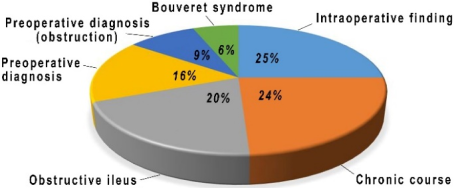
In the chronic phase of its existence, as a rule, patients indicate weight loss, various signs of dyspepsia, abundant fatty feces. In total, such development of the disease was revealed in 13 (24%) patients (Figure 1).
Figure 1. The main manifestations of cholecystodigestive fistula,%.
However, only in 9 (16%) patients was the presence of a cholecystodigestive fistula suspected before surgery. In 14 (25%) patients, such a fistula was an intraoperative finding. Another manifestation of a cholecystodigestive fistula may be signs of gastric outlet stenosis, which was observed by the authors in 3 (6%) patients with Bouveret syndrome, i.e., when a duodenal stone obstructed the duodenum at the level of its bulb. Another 11 (20%) patients developed biliary obstruction due to a cholecystodigestive fistula. It should be noted that only 5 (9%) patients were diagnosed before surgery. This is due to the absence of a history of biliary disease and specific symptoms of obstruction in the early stages of its development. It is only when the stone is fixed in the ileum that the picture of acute intestinal obstruction unfolds, which usually occurs by the third to tenth day of the disease. This development of the complication confirms the opinion of most surgeons that operations for biliary obstruction are often delayed, and its nature is only recognized during surgery. The main signs of a "shrunken" gallbladder were manifestations of acute or chronic cholecystitis, and the key symptoms of the Miriszi syndrome included jaundice and cholangitis.
As can be seen from the above, the manifestations of rare complications of cholelithiasis are nonspecific, and the focus of their recognition is shifted to special research methods, the significance of which is also ambiguous. The easiest way to diagnose a spontaneous external bile fistula is through fistulography, which not only confirms the presence of a fistula but also often reveals its main cause, which is often cholelithiasis.
Another method that is quite informative and suggests a rare complication is ultrasound, which allows us to see large stones and sometimes their protrusion into the dilated hepaticocholedoch in the case of Miriszi syndrome. The most revealing ultrasound findings in this syndrome were the simultaneous detection of a "shrunken" gallbladder and a large cholelithiasis, which is highly characteristic of Miriszi syndrome. The final diagnosis of Miriszi syndrome can only be made by direct contrasting of the bile ducts, which is most easily achieved by endoscopic retrograde cholangiography or magnetic resonance pancreatocholangiography.
A very demonstrative, necessary, and at the same time quite traditional procedure is an abdominal X-ray in case of biliary obstruction. The detection of a triad using this method - 1) aerobilia, 2) signs of small bowel obstruction, and 3) a shadow of a calculus in the small intestine - clearly indicated the cause of the patient's suffering.
A very demonstrative, necessary, and at the same time quite traditional procedure is an abdominal X-ray scan in cases of biliary obstruction. The detection of a triad using this method - 1) aerobilia, 2) signs of small bowel obstruction, and 3) a shadow of a calculus in the small intestine - clearly indicated the cause of the patient's suffering.
Also informative is the radiocontrast study of the passage of barium suspension through the small intestine, revealing a cholecystodigestive fistula, levels and arcades of the small intestine. A more demonstrative picture showing signs of obstruction and the presence of cholelithiasis and a stone obstructing the small intestine is revealed by computer tomography.
As noted, the course of rare complications of cholelithiasis is accompanied by a mild-symptom picture and sometimes occurs under the guise of a tumor, in particular, of the pancreas, duodenal ulcer disease. In two such cases, a Bouvet syndrome was detected during computed tomography, characterized by obstruction at the duodenal bulb level and symptoms of duodenal obstruction. In another case, a cholecystoduodenal fistula was mistaken for a postbulbar ulcer with duodenal stenosis during gastroduodenoscopy. However, a large stone was not detected distal to the stenosis.
It should be emphasized that, despite the high information content of special research methods, unfortunately, not all of them are feasible in urgent surgery, when an operation is necessary on an urgent or emergency basis, and the data obtained even in the same type of rare complications of cholelithiasis are variable, which makes their interpretation difficult. Therefore, the sequence of studies, their necessity, and sufficiency are determined for each individual patient. In general, as the conducted studies showed, in 57 (61%) patients, a rare complication of cholelithiasis turned out to be an intraoperative finding.
Observations of spontaneous external bile fistulas are extremely rare. So, foreign surgeons have recorded only 100 such cases over the past century . The authors of this study observed 5 such patients, and in 3 of them, a combination of complications of cholelithiasis was detected, such as Miriszi syndrome, large choledocholithiasis, and a "shrunken" gallbladder, which was a consequence of stone carriage for 13, 17, and 23 years in elderly and senile patients with concomitant diseases, and they were denied surgical treatment on this basis. Two other patients underwent irrational surgical tactics aimed at relieving acute cholecystitis using a cholecystostomy, and after its removal, they were observed for 5 and 7 years, respectively, without undergoing subsequent surgery.
We believe that the most reasonable approach to acute cholecystitis is to operate on all patients with acute cholecystitis, and the timing of the operation and the type of treatment depend solely on the degree of destruction of the gallbladder and the involvement of the "gallbladder - biliary ducts - pancreas" system in the disease process.
Regarding spontaneous external bile fistulas, we consider "open" cholecystectomy to be the optimal type of intervention, with the possibility of expanding the treatment if a combination of complications is detected. Of the 32 patients with internal fistulas, a complex variant was found in 2 patients, who simultaneously had a bilobiliary fistula (Mirizi syndrome) and a cholecystoduodenal fistula. In addition, 11 patients with a cholecystoduodenal fistula developed biliary obstruction due to gallstones, and 3 patients developed duodenal obstruction due to a large gallstone (Bouvet syndrome).
The main operation for cholecystodigestive fistulas is their disconnection, cholecystectomy, and suture of the fistula. In the case of a fistula with the colon, a two-barrel transversostomy was performed on the anterior abdominal wall. Finally, in 2 cases of a combination of a cholecystoduodenal fistula and Mirizzi syndrome, the fistula was dissected, the duodenal fistula was sutured, and a cholecystectomy and T-drainage of the bile ducts were performed.
We believe that in some cases of cholecystoduodenal fistulas, after dissection, it is possible to close the duodenal fistula using a "plug" at the level of the pylorus and a gastroenteroanastomosis or a Roux-en-Y gastric resection. In cases where cholelithiasis in Miriszi syndrome cannot be resolved using endoscopic papillotomy, it is necessary to perform a biliary anastomosis. We do not recommend using fistulas for drainage and biliodigestive anastomosis due to the risk of failure. However, in one case, this rule was ignored due to the patient's critical condition, resulting in a successful treatment outcome.
Intestinal obstructive cholelithiasis is closely related to cholecystodigestive fistulas, which is a continuation of the development of fistulas and a sign of an increase in the number and quality of complications. This type of complication occurs when large stones migrate from the gallbladder, most often into the duodenum and then into the small intestine, obstructing the ileum at its narrowest point.
The principal feature of the clinical picture of obstructive biliary obstruction is its long-term development, intermittent and nonspecific manifestations, delayed recognition, and, as a result, late surgery. These circumstances lead to severe water and electrolyte imbalance and high mortality among such patients.
A similar course of the disease was revealed in 11 patients, of whom 4 died. A retrospective analysis, the accumulation of experience in the treatment, alertness to the possibility of occurrence of this severe complication served to the fact that the rest of the noted number of patients were successfully treated.
The main condition for achieving encouraging results in the treatment of a combination of internal bile fistulas and biliary stone obstruction is considered to be the reduction of the period of observation and the performance of timely and adequate surgery. We believe that in emergency surgery, it is justified to perform combined operations aimed at curing cholelithiasis and resolving obstructive ileus. Otherwise, as was the case in one of the observations, stones that caused the recurrence of ileus would continue to migrate from the retained gallbladder. Additionally, if the gallbladder is retained, there is a risk of acute cholecystitis occurring in the early stages after surgery, unless a cholecystectomy or, as a last resort, a cholecystolithotomy with cholecystostomy has been performed.
If it is not possible to perform a cholecystectomy at the same time due to local inflammatory changes, it is important to ensure that there are no stones remaining in the gallbladder. In such cases, it is advisable to perform a cholecystectomy as a second step (2-3 months later), which was done in one patient. Based on the clinic's experience, the use of A.V. Vishnevsky's modified mucoclasia of the gallbladder or the resection of the gallbladder according to the classification of foreign surgeons can be considered for "difficult" cases of cholecystectomy.
In addition, it has been confirmed that positive treatment results can be achieved using long-term nasojejunal intubation for decompression, lavage, intestinal decontamination, and enteral nutrition. These measures reliably control and correct the resulting disorders of the motor-evacuatory and absorptive functions of the intestine, which is essential for successful treatment of patients with intestinal obstruction.
However, it is much easier to prevent the development of rare complications of cholelithiasis, which can be achieved by implementing a modern approach to the treatment of cholelithiasis, which focuses on early, minimally invasive surgery during the cold period of the disease, regardless of age, and after the compensation of concomitant diseases. This approach eliminates the need for surgery, as was observed in a case of obstructive cholelithiasis-related small bowel obstruction, Miriszi syndrome, and choledochoduodenal fistula, 57 years after the diagnosis of cholelithiasis.
Another complication, the "shrunken" gallbladder, as a late manifestation of long-standing cholelithiasis, also presents a rather difficult problem for the surgeon, both in terms of diagnosis and surgery. Eleven such patients were studied, with an average disease duration of 11 years, and four patients had not previously undergone surgical treatment.
Of course, the detection of a reduced-sized, thick-walled, and sometimes non-lumenous gallbladder during an ultrasound examination or computed tomography scan suggests that it may be a sign of malignant transformation in the context of cholelithiasis. We believe that both gallbladder cancer and benign "shrinking" of the gallbladder require surgical treatment, the nature of which depends on the intraoperative findings and urgent histological examination. When the intraoperative confirmation of benign shrinkage of the gallbladder, the surgeon faces a variety of questions related to the safe removal of the gallbladder, mainly to prevent injury to the extrahepatic bile ducts and major vessels. To this end, separate techniques are proposed, the essence of which is to "get into the layer" when isolating the gallbladder and "carefully and slowly" operate.
In addition to the well-known technique of removing an inflamed or "shrunken" gallbladder after evacuating the bile, opening the bladder in a longitudinal direction, removing the stones, and performing a "finger" cholecystectomy, it is advisable to perform a subserous gallbladder removal or a mucoclasia modification by A. V. Vishnevsky.
The essence of the first method is to enter under the serous membrane of the gallbladder after hydraulic preparation and remove it together with perifocal inflammatory tissues from the mucosa of the gallbladder and the cystic duct, perform cholecystectomy and ligate the cystic duct or introduce a drainage of the hepaticocholedoch through it. When performing mucoclasia of the gallbladder in the modification of A. V. Vishnevsky, the differentiated walls of the gallbladder are removed, bleeding areas are stitched, and the remaining mucosa is electro-destructed.
Something similar is performed by foreign surgeons during laparoscopic cholecystectomy, calling this type of surgery "partial resection of the gallbladder" . The latter point of view contradicts the opinion of the authors of this article. We believe that the detection of a "shrunken" gallbladder is a contraindication for performing laparoscopic cholecystectomy.
In the problem of the Mirizzi syndrome, the concept of the syndrome itself and the choice of surgical treatment remain the most controversial. According to Pablo Mirizzi (1948), this syndrome is characterized by obstruction of the common bile duct (sindrome del conducto hepatico) . It is caused by the destruction of the cystic duct and the neck of the gallbladder as a result of acute cholecystitis, the prolapse of a stone into the hepaticocholedochus, and the development of obstructive jaundice. It is evident that the main substrate of this syndrome is a cholecystocholedochal fistula. Recently, a number of surgeons have identified five types of the syndrome, which do not fully correspond to the published original source, and the first type does not have anything to do with the Mirizzi syndrome and is an extrabiliary compression of the common hepatic duct described by Ker in 1905 . The other 4 types of the syndrome are an indicator of the degree of stone prolapse into the bile ducts or a combination of the Mirizi syndrome with a cholecystoduodenal fistula, which are essentially stages of the syndrome's progression, and the degree of destruction of the hepaticocholedoch affects the choice of the method for completing the surgical procedure.
As shown by the observations of 47 patients with Mirizzi syndrome, this condition was recognized only in 14 patients in the preoperative period. Attention was drawn to the less pronounced local changes during surgery outside the phase of exacerbation of calculous cholecystitis, when there is no jaundice or cholangitis. In such cases, the surgery is more anatomical, there are fewer non-standard situations, and the tissues and structures, especially the hepatoduodenal ligament, are clearly visible, resulting in better surgical outcomes. The cause of death (7 deaths, or 15% of all patients with Miriszi syndrome) was obstruction of the bile ducts, "toxic" cholangitis, or cholangiogenic abscesses, a combination of several complications, and delayed surgical intervention.
5. Recommendations
The authors' proposals for rational surgical treatment of Mirizzi syndrome are as follows:
1) excision of the gallbladder with leaving 1 cm of a cholecystodigestive fistula on the hepaticocholedochus;
2) electrocoagulation of the mucous membrane of the remaining part of the fistula;
3) revision of the bile ducts through the fistula is possible only if the fistula is connected to the hepaticocholedochus at a right angle;
4) in most cases, after electrocoagulation of the mucous membrane, the fistula is sutured with separate atraumatic sutures and a cholecystectomy is performed below or above the fistula;
5) we do not use the fistula for creating biliary anastomoses and draining the choledochus;
6) we use the choledochotomy opening to create biliary anastomoses and drain the choledochus due to the risk of suture failure in the area of inflammatory changes in the fistula;
7) large defects of the hepaticocholedochus are sutured with the demucated part of the posterior wall of the gallbladder.
These recommendations for Miriszi syndrome are not final and may vary depending on the patient's overall condition, the specific situation, and any local changes in the surgical area that the surgeon may encounter during the operation.
6. Conclusions
The main reason for the development of rare complications of cholelithiasis is a long-term and/or asymptomatic course of the disease. Often, a long-term course of the disease is caused by an unjustified refusal to perform surgery due to the patient's age, the presence of concomitant diseases, and the risk of general anesthesia. The best way to prevent rare complications of cholelithiasis is to perform timely surgery during the cold period, preferably using minimally invasive techniques.
Recognizing rare complications of cholelithiasis can be challenging, as they may occur in combination. Due to the development of a combination of complications of cholelithiasis and decompensation of concomitant diseases, surgical interventions are often delayed. This is the main cause of fatal outcomes.
The developed strategy for the surgical treatment of rare complications of cholelithiasis, aimed at early recognition and surgical intervention in such patients, resulted in a favorable outcome in the immediate postoperative period in 85 patients. Eleven patients died, and the overall mortality rate was 11.6%.
Author Contributions
Vladimir Glabay: Conceptualization, Investigation, Resources, Supervision, Validation
Vitaliy Tsvetkov: Methodology, Project administration, Validation Writing – original draft
Rashid Askerkhanov: Data curation, Formal Analysis, Writing - review & editing
Zaira Eldarova: Visualization, Writing – original draft, Writing – review & editing
Vakhtang Gobedzhishvili: Data curation, Investigation, Software
Data Availability Statement
The data is available from the corresponding author upon reasonable request.
Conflicts of Interest
The authors declare no conflicts of interest.
References
[1] Clavien, P. A. Baillie, J. Diseases of the Gallbladder and Bile Ducts: diagnosis and treatment. Blackwell scince. Malden, Massachusetts, 2001. 346 p.
[2] Acute calculous cholecystitis. New information on cholelithiasis. A practical guide. Editors: Buriev, I. M., Melkonyan, G. G. Moscow: GEOTAR-Media, 2020. 272 с.
[3] Glabay, V. P., Eldarova, Z. E. Rational Approach to Surgical Treatment of Acute Biliary Pancreatitis. Surgery Eastern Europe. 2024; 13(4): 512-521.
[4] Natroshvili, I. G. Ways to optimize treatment tactics for patients with acute calculous cholecystitis. Results of a multicenter study [dissertation]. Yekaterinburg: Ural State Medical University; 2019. 214 p.
[5] Chinikov, M. A. Treatment of patients with calculous cholecystitis with a 24-hour hospital stay [dissertation]. Moscow: Peoples' Friendship University of Russia; 2019. 255 p.
[6] Glabay, V. P., Askerkhanov, R. G., & Matveeva, G. K. Surgical Management of Patients with Rare Complications of Cholelithiasis. Annals of Surgical Hepatology. 2009; 14(1): 57-63.
[7] Galperin, E. I., Akhaladze, G. G., Kotovsky, A. E., Ungurjanu, T. V., Chevokin, A. Yu. Mirizzy Syndrome: Peculiarities of Diagnoses and Management. Annals of Surgical Hepatology. 2006; 11(3): 7-10.
[8] Аrdasenov T. B., Budzinsky S. A., Pankov А. G., Bachurin А. N., Shapovalyants S. G. Peculiarity of Surgical Management of Difficult Common Bile Duct Stones of Difficult Common Bile Duct Stones. Annals of HPB Surgery. 2013; 18(1): 23-28.
[9] Glabay, V. P., Tsvetkov, V. O., Gobedzhishvili, V. V., Eldarova, Z. E., Lesovik, V. S., Orlov B. B. Radical treatment of unformed intestinal fistulas. Medical News of North Caucasus. 2022; 17(2): 199-201.
[10] Askerkhanov, R. G. Rare complications of cholelithiasis and their surgical treatment [dissertation]. Moscow: A. V. Vishnevsky Institute of Surgery; 2008. 103 p.
[11] Vasilchenko, M. I., Ratchyk, V. M., Eldarova, Z. E. Instrumental and laboratory diagnostics of early complications in acute pancreatitis. Experimental and Clinical Gastroenterology. 2022; 8: 106-112.
[12] Strasberg, S. M., Pucci, M. J., Brunt, L. M., Deziel, D. J. Subtotal Cholecystectomy - «Fenestrating» vs «Reconstituting». Journal of the American College of Surgeons. 2016; 222(1): 89-96.
[13] Lunevicius, R. Review of the Literature on Partial Resections of the Gallbladder, 1898-2022: The Outline of the Conception of Subtotal Cholecystectomy and a Suggestion to Use the Terms «Subtotal Open-Tract Cholecystectomy» and «Subtotal Closed-Tract Cholecystectomy». J Clin Med. 2023 Feb 3; 12(3): 1230.
[14] Ramírez-Giraldo, C., Torres-Cuellar, A., Van-Londoño, I. State of the art in subtotal cholecystectomy: An overview. Frontiers in Surgery. 2023 Apr 21; 10: 1142579.
[15] Mirizzi, P. L. Sindrome del conducto hepático. Journal International de Chirurgie. 1948; 8(3): 731-777.
Cite This Article
  • APA Style

    Glabay, V., Tsvetkov, V., Askerkhanov, R., Eldarova, Z., Gobedzhishvili, V. (2026). Rational Surgical Management of Rare Complications of Cholelithiasis. International Journal of Clinical and Experimental Medical Sciences, 12(3), 57-63. https://doi.org/10.11648/j.ijcems.20261203.12

    Copy | Download

    ACS Style

    Glabay, V.; Tsvetkov, V.; Askerkhanov, R.; Eldarova, Z.; Gobedzhishvili, V. Rational Surgical Management of Rare Complications of Cholelithiasis. Int. J. Clin. Exp. Med. Sci. 2026, 12(3), 57-63. doi: 10.11648/j.ijcems.20261203.12

    Copy | Download

    AMA Style

    Glabay V, Tsvetkov V, Askerkhanov R, Eldarova Z, Gobedzhishvili V. Rational Surgical Management of Rare Complications of Cholelithiasis. Int J Clin Exp Med Sci. 2026;12(3):57-63. doi: 10.11648/j.ijcems.20261203.12

    Copy | Download

  • @article{10.11648/j.ijcems.20261203.12,
      author = {Vladimir Glabay and Vitaliy Tsvetkov and Rashid Askerkhanov and Zaira Eldarova and Vakhtang Gobedzhishvili},
      title = {Rational Surgical Management of Rare Complications of Cholelithiasis},
      journal = {International Journal of Clinical and Experimental Medical Sciences},
      volume = {12},
      number = {3},
      pages = {57-63},
      doi = {10.11648/j.ijcems.20261203.12},
      url = {https://doi.org/10.11648/j.ijcems.20261203.12},
      eprint = {https://article.sciencepublishinggroup.com/pdf/10.11648.j.ijcems.20261203.12},
      abstract = {Most patients with gallstone disease undergo elective surgery. However, some patients do not undergo surgery because, despite long-term treatment, they experience only rare complications, the incidence of which does not exceed 1.4%. Rare complications of gallstone disease include external biliary fistulas, cholecystocholedocheal fistulas or Mirizzi syndrome, micro or "large" choledocholithiasis, "shrunken" gallbladder, gallbladder calcification (porcelain gallbladder), and others. Recognizing these rare complications of gallstone disease presents significant challenges, and various types of imaging are the primary methods for their detection. Development of rational surgical management to improve treatment outcomes of rare complications of cholelithiasis. A total of 5,449 patient records for cholelithiasis were reviewed. Of these, 3,332 (61%) were admitted for emergency indications and 2,117 (39%) for elective surgery. Of all patients with calculous cholecystitis, 96 had rare complications, representing a 1.74% incidence. The male-to-female ratio was 1: 4, and the average age of the patients was 70 years. A review of the anamnestic data revealed that the patients had suffered from cholelithiasis for between 5 and 57 years. Of the 96 patients operated on for rare complications of cholelithiasis, 16 (17%) were operated on for emergency indications with acute cholecystitis, 30 (32%) due to the ineffectiveness of conservative treatment, and 14 (15%) due to the failure of endoscopic papillotomy undertaken to resolve mechanical jaundice and cholangitis or due to the development of obstructive cholelithiasis. A study of clinical observations of patients with rare complications of gallstone disease revealed the absence of any pathognomonic manifestations. Only in cases of spontaneous external biliary fistulas, with bile leaking onto the anterior abdominal wall, was the diagnosis immediately apparent. The primary surgical procedure for cholecystodigestive fistulas is their dissection, cholecystectomy, and fistula closure. In cases where choledocholithiasis associated with Mirizzi syndrome cannot be resolved with endoscopic papillotomy, a biliary anastomosis is necessary. The developed surgical approach for rare complications of cholelithiasis aimed at early recognition and prompt surgical intervention led to a favorable short-term outcome in 85 of 96 patients. The overall mortality rate was 11.6% (n=11). The main cause of rare complications of cholelithiasis is a prolonged and/or asymptomatic course of the disease. The best method for preventing this disease is timely surgery during the cold period, preferably minimally invasive. Decompensation of concomitant diseases against the background of a combination of complications of cholelithiasis primarily leads to a fatal outcome.},
     year = {2026}
    }
    

    Copy | Download

  • TY  - JOUR
    T1  - Rational Surgical Management of Rare Complications of Cholelithiasis
    AU  - Vladimir Glabay
    AU  - Vitaliy Tsvetkov
    AU  - Rashid Askerkhanov
    AU  - Zaira Eldarova
    AU  - Vakhtang Gobedzhishvili
    Y1  - 2026/08/18
    PY  - 2026
    N1  - https://doi.org/10.11648/j.ijcems.20261203.12
    DO  - 10.11648/j.ijcems.20261203.12
    T2  - International Journal of Clinical and Experimental Medical Sciences
    JF  - International Journal of Clinical and Experimental Medical Sciences
    JO  - International Journal of Clinical and Experimental Medical Sciences
    SP  - 57
    EP  - 63
    PB  - Science Publishing Group
    SN  - 2469-8032
    UR  - https://doi.org/10.11648/j.ijcems.20261203.12
    AB  - Most patients with gallstone disease undergo elective surgery. However, some patients do not undergo surgery because, despite long-term treatment, they experience only rare complications, the incidence of which does not exceed 1.4%. Rare complications of gallstone disease include external biliary fistulas, cholecystocholedocheal fistulas or Mirizzi syndrome, micro or "large" choledocholithiasis, "shrunken" gallbladder, gallbladder calcification (porcelain gallbladder), and others. Recognizing these rare complications of gallstone disease presents significant challenges, and various types of imaging are the primary methods for their detection. Development of rational surgical management to improve treatment outcomes of rare complications of cholelithiasis. A total of 5,449 patient records for cholelithiasis were reviewed. Of these, 3,332 (61%) were admitted for emergency indications and 2,117 (39%) for elective surgery. Of all patients with calculous cholecystitis, 96 had rare complications, representing a 1.74% incidence. The male-to-female ratio was 1: 4, and the average age of the patients was 70 years. A review of the anamnestic data revealed that the patients had suffered from cholelithiasis for between 5 and 57 years. Of the 96 patients operated on for rare complications of cholelithiasis, 16 (17%) were operated on for emergency indications with acute cholecystitis, 30 (32%) due to the ineffectiveness of conservative treatment, and 14 (15%) due to the failure of endoscopic papillotomy undertaken to resolve mechanical jaundice and cholangitis or due to the development of obstructive cholelithiasis. A study of clinical observations of patients with rare complications of gallstone disease revealed the absence of any pathognomonic manifestations. Only in cases of spontaneous external biliary fistulas, with bile leaking onto the anterior abdominal wall, was the diagnosis immediately apparent. The primary surgical procedure for cholecystodigestive fistulas is their dissection, cholecystectomy, and fistula closure. In cases where choledocholithiasis associated with Mirizzi syndrome cannot be resolved with endoscopic papillotomy, a biliary anastomosis is necessary. The developed surgical approach for rare complications of cholelithiasis aimed at early recognition and prompt surgical intervention led to a favorable short-term outcome in 85 of 96 patients. The overall mortality rate was 11.6% (n=11). The main cause of rare complications of cholelithiasis is a prolonged and/or asymptomatic course of the disease. The best method for preventing this disease is timely surgery during the cold period, preferably minimally invasive. Decompensation of concomitant diseases against the background of a combination of complications of cholelithiasis primarily leads to a fatal outcome.
    VL  - 12
    IS  - 3
    ER  - 

    Copy | Download

Author Information