Research Article | | Peer-Reviewed

Clinical Profile and Etiological Spectrum of Exudative Pleural Effusion

Received: 8 August 2026     Accepted: 24 August 2026     Published: 18 September 2026
Views:       Downloads:
Abstract

Background: Exudative pleural effusion is commonly caused by tuberculosis and malignancies, often presenting significant diagnostic challenges in clinical practice. The need to combine clinical characteristics with a cost effective and targeted approach towards making a definitive diagnosis cannot be overemphasized. This study examines the clinical presentation, demographic characteristics, and etiological spectrum of exudative pleural effusion in a Nigerian tertiary care setting. Methods: This prospective study involved 58 patients, aged 19 to 87 years who were referred to the cardiothoracic unit of the University of Benin Teaching Hospital over a period of one year with undiagnosed exudative pleural effusion. The demographics, clinical presentation and investigations including laboratory, radiological and pleural fluid characteristics were recorded. The data were subsequently analyzed using SPSS version 25, with results expressed as frequencies, percentages, means, and standard deviations. Results: The study revealed malignancy (50%) and tuberculosis (43.2%) as the leading causes of undiagnosed exudative pleural effusion in our environment. Dyspnea (91.4%), cough (87.9%), and chest pain (79.3%) were the most common symptoms. It also demonstrated male preponderance with a male to female ratio of 1.1:1 and a mean age of 51.16±17.487. Left sided effusion predominates (56.9%) as compared to the right (43.1%). Hemorrhagic effusion featured prominently in cases of malignancy (82.8%) while turbid effusion was commoner in cases of tuberculosis (72.0%). Conclusion: These findings highlight the need for a thorough clinical evaluation and targeted diagnostic strategies to optimize patient outcomes.

Published in International Journal of Cardiovascular and Thoracic Surgery (Volume 12, Issue 5)
DOI 10.11648/j.ijcts.20261205.12
Page(s) 122-130
Creative Commons

This is an Open Access article, distributed under the terms of the Creative Commons Attribution 4.0 International License (http://creativecommons.org/licenses/by/4.0/), which permits unrestricted use, distribution and reproduction in any medium or format, provided the original work is properly cited.

Copyright

Copyright © The Author(s), 2026. Published by Science Publishing Group

Keywords

Exudative Pleural Effusion, Tuberculosis, Lung Cancer, Clinical Presentation, Ultrasonography, Pleural Biopsy

1. Introduction
Pleural effusion, characterized by abnormal fluid accumulation in the pleural cavity, is a diagnostic challenge due to its varied etiology and thus requires comprehensive evaluation to effectively determine the specific cause in order to achieve successful management . Many common medical conditions manifest as pleural effusion but a vast majority of them are of benign nature . It has however been documented that tuberculosis and malignancies are the most frequent causes of exudative pleural effusion worldwide , with geographical and socio-economic factors influencing prevalence. Pleural fluid collections are either transudative or exudative. The Light's criteria provides the basis for differentiating transudative effusion from exudative pleural fluid collections. Pleural effusion is exudative if it satisfies any one of the following criteria: pleural fluid protein/serum protein ratio >0.5 or pleural fluid lactate dehydrogenase (LDH)/serum LDH ratio >0.6 or pleural fluid LDH level greater than two-thirds the upper limit of normal .
The initial diagnostic evaluation of pleural effusion includes chemical, microbiological studies and cytological analysis . Approximately 40% of pleural effusions remain undiagnosed after an initial thoracocentesis thereby necessitating pleural biopsy procedure. The determination of the etiology of pleural effusion is an important factor for effective treatment . The complex interplay of clinical presentation, patient history, and physical examination forms the basis for understanding the etiological landscape of pleural fluid collection .
Pleural effusion is seen on chest radiograph as an area of homogenous opacity with blunting of the costophrenic and cardiophrenic angles. In cases where the fluid in the pleural cavity is small, a lateral view of the chest X-ray gives a better imaging than a posterolateral one. Chest ultrasonography is gaining momentum in the recent times in the evaluation of patients with pleural effusion as it appears to be more sensitive than chest radiograph. It is safe and has no radiation but possesses the benefit of repeatability . It is an efficient tool in the hands of experts when identifying loculations and septations in the pleural space and can as well be used to guide thoracocentesis and choose the site of pleural biopsy and insertion of intercostal tube. A computerized tomography scan is usually indicated in conditions where lung parenchyma and mediastinal abnormalities are suspected. It also helps to quantify the amount of fluid in the pleural space and aids image guided biopsy.
In industrialized nations of the world the estimated prevalence of pleural effusion is 320 cases per 100,000 people, with etiological distribution related to the prevalence of the underlying diseases . In the United States the incidence of pleural effusion is estimated to be about 1.5 million cases annually whereas in the UK, more than 0.3% of the population has pleural effusion each year . It is reported that, pleural diseases account for 7.7% of respiratory morbidity in the South West Nigeria, the majority occurring in patients between the ages of 15 and 44 years. Ogunleye et al in their work on aetiology and demographic attributes of common pleural collections in an African population reported 372 cases of pleural effusion over a 55-month period with male to female ratio of 1:1 and most of them between the ages of 20 and 49 years .
The objectives of the study includes
To determine the aetiology of undiagnosed exudative pleural effusion referred to the cardiothoracic unit of the University of Benin Teaching Hospital.
To study the clinical presentation of the patients with undiagnosed exudative pleural effusion.
To determine the correlation between the nature of effluents from the pleural space and the final diagnosis.
2. Method
2.1. Study Setting
The study was conducted in the Cardiothoracic Unit, Department of Surgery, University of Benin Teaching Hospital (UBTH), a tertiary healthcare centre in southern Nigeria. The hospital serves as a major referral centre for patients with thoracic diseases from Edo State and neighbouring states.
2.2. Study Design and Study Population
This was a prospective observational study conducted over a period of one year. The study population comprised adult patients referred to the Cardiothoracic unit with undiagnosed exudative pleural effusion.
2.3. Participant Eligibility
Patients were eligible for inclusion if they were aged 18 years or older and had clinical, radiological, and laboratory evidence of exudative pleural effusion according to Light's criteria. Patients who did not meet the criteria for exudative pleural effusion or from whom adequate clinical and diagnostic data could not be obtained were excluded.
2.4. Sample Size and Sampling
A total of 58 consecutive patients who met the eligibility criteria were enrolled during the study period.
2.5. Clinical and Demographic Data Collection
Demographic and clinical information was obtained using a structured data collection instrument. Variables collected included age, sex, presenting symptoms, relevant clinical findings, and other pertinent clinical characteristics. Results of relevant laboratory and radiological investigations were also recorded.
2.6. Pleural Fluid Evaluation
Pleural fluid obtained from eligible participants was subjected to appropriate laboratory investigations for the evaluation of the underlying cause of the effusion. Pleural fluid characteristics and relevant biochemical, microbiological, and cytological findings were documented.Exudative pleural effusion was classified according to Light's criteria.
2.7. Cytological and Histopathological Evaluation
Pleural fluid cytological examination was performed where indicated. Pleural tissue obtained through biopsy was submitted for histopathological examination. Cytological and histopathological findings were used, along with the clinical, radiological, and laboratory findings, to establish the definitive aetiology of the pleural effusion.
2.8. Definition of Final Diagnosis
The final aetiological diagnosis was established based on the overall clinical assessment and available diagnostic findings, including pleural fluid investigations, cytology, and histopathological examination.
2.9. Data Management and Statistical Analysis
Data were entered, coded, and analysed using the Statistical Package for the Social Sciences (SPSS), version 25. Categorical variables were summarized using frequencies and percentages. Continuous variables were expressed as means and standard deviations, as appropriate.
2.10. Ethical Considerations
Ethical approval was obtained from the appropriate institutional research ethics committee before commencement of the study. Written informed consent was obtained from all participants before enrolment. Patient information was treated confidentially and used solely for the purposes of the study.
3. Results
Demographics: The study included 30 males and 28 females, with a male-to-female ratio of 1.1:1. The mean age was 51.16 ± 17.49 years.
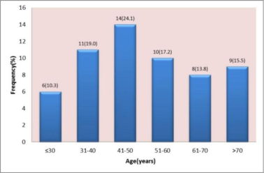
Figure 1. Age distribution of the patients.
Figure 1 shows the bar chart of age distribution of the patients in the study. The age range of patients was between 19-87 years. The peak age distribution was 41-50 years accounting for 24.1% of the total population. The mean age of the study population was 51.16±17.487. Over 40% of the population was between the ages of 41 to 60 years while 29.3% each were below 40 years and above 60 years respectively.
Table 1. Showing the Clinical Features of the patients.

Symptom

Frequency n=58

Percentage (%)

Dyspnoea

53

91.4

Cough

51

87.9

Chest Pain

46

79.3

Haemoptysis

25

43.1

Weight Loss

34

58.6

Fever

10

17.2

Anorexia

8

13.8

Clinical Presentation The most common symptoms were dyspnea (91.4%), cough (87.9%), and chest pain (79.3%). Weight loss (58.6%) and hemoptysis (43.1%) were also observed (Table 1).
Table 2. Showing the Causes.

Diagnosis

Frequency n=58

Percentage (%)

Malignancy

29

50.0

Tuberculosis

25

43.2

Empyema Thoracis

2

3.4

Thoracic Endometriosis

2

3.4

Etiology: Malignancy accounted for 50% of cases, followed by tuberculosis at 43.2%. Empyema thoracis and thoracic endometriosis were rare (Table 3).
Table 3. Pleural Fluid Characteristics.

Nature of Effluents

Malignancy (%)

Tuberculosis (%)

Empyema (%)

Total

Haemorrhagic

82.8

20.0

0.0

Turbid

10.3

72.0

0.0

Purulent

6.9

8.0

100

Hemorrhagic effusion predominated in malignancy (82.8%), while turbid effusion was commoner in tuberculosis (72.0%) (Table 4).
Table 4. Mean and Standard Deviation of Protein and LDH Investigations.

Range

Mean

Std. Deviation

Protein

Pleural Fluid

2.90 (3.2 – 6.1)

4.20

0.690

Serum

2.80 (5.1 – 7.9)

6.14

0.746

Ratio

0.23 (0.59 – 0.82)

0.68

0.061

LDH

Pleural Fluid

636 (210 – 846)

436.28

149.445

Serum

392 (228 – 620)

332.07

73.513

Ratio

0.95 (0.79 – 1.74)

1.30

0.255

The above table shows that all the patients that participated in the study had exudative pleural effusion. The figures above satisfied light’s criteria for exudative effusion; the ratio of pleural fluid protein to serum protein was greater than 0.5 and the ratio of pleural fluid LDH to serum LDH was greater than 0.6 for all participants. For all cases studied, the absolute value of pleural fluid protein and pleural fluid LDH were greater than 3g/dl and 200IU/l respectively.
Table 5. Clinical Investigations.

Variables

Frequency (n = 58)

Percent

Extent of Effusion

Massive pleural effusion

26

44.8

Moderate pleural effusion

32

55.2

Side of Effusion

Left sided pleural effusion

33

56.9

Right sided pleural effusion

25

43.1

Full Blood Count

Normal

40

69.0

Leucocytosis

7

12.0

Lymphocytosis

11

19.0

Table 5 shows the results of chest X-ray and the full blood count parameters for all the patients in the study. It demonstrates that 44.8% of the cases presented with massive pleural effusion while 55.2% came with moderate pleural effusion. The effusions were left sided in 56.9% and right sided in 43.1%. The full blood count shows values within normal range in 69% of cases. However 12% of cases had leukocytosis while 19% had lymphocytosis.
4. Discussion
This study was designed to evaluate the etiological spectrum and the clinical profile of patients with exudative pleural effusion referred to the cardiothoracic unit of the University of Benin Teaching Hospital. The study confirms malignancy and tuberculosis as the leading causes of exudative pleural effusion in our setting.
4.1. Socio-demographic Characteristics
Over a period of one year a total number of 58 consecutive patients with laboratory evidence of pleural effusion were recruited into the study. Thirty (30) were males while 28 were females with a male to female ratio of 1.1: 1. These findings are in keeping with the observations noted in other studies that have reported male preponderance in patient with pleural diseases. This may be due to the high prevalence of tobacco smoking and alcohol consumption in males compared to females in our environment. The mean age for the participants in this study was 51.16±17.487 and their ages range from 19 to 87 years. Previous studies have documented similar figure for mean age and age range with slight disparities. A. H Diacon et al recorded a mean age range of 56 years (range 18-80), B. Heidari et al recorded a mean age of 57±17 years with a range (12-82) while A. K. Singh and colleagues in 2024 described a mean age of 48.83±22.87 with the age range of between 8 and 94 years. The mean age in patient with malignancy (61.33±14.497) in this study was higher than the mean age of patients with tuberculosis (42.04±14.616) and other conditions. This is consistent with the findings in a work done by B. Heidari et al in their study on exudative pleural effusion: effectiveness of pleural fluid analysis and pleural biopsy . A similar observation was made in another study on the etiology of exudative pleural effusion among adults: differentiating between tuberculous and other causes, a multicenter prospective cohort study in which the mean age of patient with malignancy (58.33±13.26) was higher than that in tuberculosis (37.78±10.91).
4.2. Mode of Presentation
The common symptoms presented by participants in this study were dyspnoea, 53 (91.4%), cough 51 (87.9%) chest pain 46 (79.3%) and hemoptysis 25 (43.1%) of cases. Other were weight loss 34 (58.6%), fever 10 (17.2%) and anorexia 8 (13.8%). This observation is consistent with the findings from previous studies, Hoda MB et al (2013) found that the common symptoms of patients with pleural diseases in their study were dyspnoea (90.0%), cough (83.3%), and chest pains (66.7%). B. Heidari and colleagues in their study on exudative pleural effusion also documented dyspnoea, cough and chest pains as the main presenting symptoms in patients with tuberculosis and malignancy. Similarly, in agreement with this study, Sahn et al (1991) found dyspnoea and chest pains as the most common presenting symptoms in patient with pleura effusion. This is more so in patients having moderate to massive pleural effusion. Pleuritic chest pain is a cardinal presentation in these patients and it is the result of pleural inflammation. In consonance with our findings, Dhital et al, in 2009 in their assessment of the clinical profile of patients with pleural effusion found shortness of breath (83%), cough (67%) and fever (66%) as the most common clinical symptoms in their patients with pleural diseases . Adeoye PO et al (2017) found dyspnea, cough and fever as the commonest presentation in their study on etiology, clinical characteristics, and management of pleural effusion in Ilorin, Nigeria and . Laxma RS et al (2019) and Mrinal KS and colleagues (2024) found dyspnoea, cough and fever as the commonest symptoms in their studies on Clinical and Etiological Profile of an Exudative Pleural Effusion in a Tertiary Care Center and Etiological profile of Pleural Effusion: A single Centre Study. respectively.
In the last four studies highlighted, it is observed that fever featured prominently in their work compared to our index study. This may be attributed to the differences in study population and the prevalence of the predisposing and/or aetiological factors for pleural effusion across the various regions. Parapneumonic effusion featured commonly in their studies which was not the case in this work.
4.3. Aetiology
The result of this study revealed that malignancy was the leading cause of exudative pleural effusion in this group of patients referred to the cardiothoracic unit as it accounted for 50% of cases studied while tuberculosis was seen in 43.2%. These findings attest that pleural biopsy is a valuable diagnostic tool for establishing the aetiology and differentiating malignant pleural effusion from tuberculous pleural effusion in patients presenting with undetermined exudative pleural effusion as all patients in this study had pleural biopsy for histology. Two studies from TB-endemic areas share similar outcome with our study as malignant diseases were also the most frequent in their series. In a similar study of patient with exudative or transudative pleural effusion in Islamic Republic of Iran, malignant diseases were also the most frequent aetiology identified. Our study contrasted with three epidemiological studies from areas endemic in tuberculosis. In those studies, tuberculosis was found to be the most common cause of exudative pleural effusion . Various factors have been adduced to influence the aetiology for exudative pleural effusion. These factors vary according to the geographical location and the population being studied. Factors that may influence or increase the risk of malignancy include age, smoking habit, and exposure to occupational or environmental risk factors while overcrowding, poverty and malnutrition are associated with increased risk of tuberculosis. .
4.4. Pleural Fluid Characteristics and Diagnosis
Available data from this study indicate that haemorrhagic effusion featured prominently in cases of malignancy than in tuberculosis or other benign conditions. While 82.8% of all cases of malignancy had haemorrhagic effusion, only 20.0% of all cases of tuberculosis had haemorrhagic effusion. It was also observed that massive effusions were more in malignant conditions than in tuberculous effusion. These observations are in keeping with findings of Heidari B et al in their study on exudative effusion. They concluded that large and bloody pleural effusions with a high protein concentration favour malignant pleural effusion rather than tuberculosis . Koppu in his descriptive study on the clinical profile of malignant pleural effusions in a tertiary care centre in 2022 also demonstrated that massive pleural effusion were commonly associated with carcinoma of the lungs . The mean and standard deviation for pleural fluid protein and LDH in our study were 4.20±0.690 and 436.2 8±149.445 respectively. This is consistent with findings in a study done by Hoda M. Bahr and colleagues in which they found 4.30±0.71 and 415.9±367.35 respectively.
In the present study, left sided effusion predominates, accounting for 56.1% of the patients with exudative pleural effusion. This figure is in consonance with findings in the study by Mrinal KS and colleagues (2024) in which they have similar observations . Our findings contrasted with the observations in a descriptive study in India that reported right sided effusion predominance . This could be attributed to the fact that while our study examined undiagnosed exudative pleural effusion, the referenced study on the other hand, described both exudative and transudative pleural effusions. Also, that the studies were conducted in two different regions of the world could be a contributory factor as the prevalence of the aetiological factors are different.
4.5. Cytology of Pleural Fluid
The diagnostic yield on cytology in cases of malignancy in the present study was 51.8%. In patients with malignant pleural effusion, cytological examination is a fast, efficient and minimally invasive procedure to establish the diagnosis. Our yield on cytology was slightly lower than the yields documented in some studies in literature as reported by NA Maskeli et al, MMkamel et al, EJ Helm et al and Cuneyt Tetikkurt et al in which they reported values between 60% and 70% However, in 2026 Ebtisan M. Gad and associates recorded a lower yield of 37.5% in their study on pleural fluid cytology versus pleural biopsy in malignant pleural effusion The discrepancy observed above may probably be due to the presence of different diseases, tumor type, tumor burden in the pleural space, tumour location, volume of pleural fluid examined, sample handling and the skills of the cytologist. Cytology of pleural fluid is the most informative and definitive initial diagnostic step in pathologic states involving the pleura . It is simple, minimally invasive, and may be considered as the best initial diagnostic tool in the hands of experienced cytologist. Also, thoracocentesis has no severe complications which makes cytology more compelling. The differential diagnosis of any pleural pathology can be considerably narrowed down by the examination of the pleural fluid alone. Cytology can lead the clinician to the definitive diagnosis or may indicate the next step that would lead to the necessary pathway to arrive at the final diagnosis. This would help to prevent unnecessary invasive interventions. In most diseases related to pleural effusion, the pleural fluid analysis yields important diagnostic information and in certain cases it provides the final diagnosis. Cytological diagnosis of malignant effusions has been reported to have yields between 40% and 87% in different studies several factors may influence its sensitivity. The effusion may develop secondary to other factors such as infection, pulmonary emboli or lymphatic blockade. The incidence of positive pleural cytology is higher in patients with a large tumor burden in the pleural surface by causing more exfoliated malignant cells to be in the effusion .
5. Conclusion
This study highlights the clinical and etiological profiles of exudative pleural effusion in a tertiary care setting. Clinical presentation varies with dyspnoea being the most common symptom. Malignancy and tuberculosis remain the most common aetiology, emphasizing the need for a thorough diagnostic approach. Incorporating imaging guidance, such as ultrasound, into routine practice can improve outcomes.
Abbreviations

LDH

Lactose Dehydrogenase

Author ContributionsAuthor Contributions
Collins Osaro Iyamu: Conceptualization, Data curation, Formal Analysis, Funding acquisition, Investigation, Methodology, Project administration, Resources, Validation, Writing – original draft, Writing – review & editing
Stanley Ukadike Okugbo: Conceptualization, Formal Analysis, Methodology, Project administration, Supervision, Validation, Writing – original draft, Writing – review & editing
Conflicts of Interest
The authors declare no conflicts of interest.
References
[1] Ferreiro L, Toubes ME, San José ME, Suárez-Antelo J, Golpe A, Valdés L. Advances in pleural effusion diagnostics. Expert Rev Respir Med. 2020 Jan; 14(1): 51-66.
[2] McLeod DT, Ternouth I, Nkanza N. Comparison of the Tru-cut biopsy needle with the Abrams punch for pleural biopsy. Thorax. 1989 Oct; 44(10): 794-6.
[3] Light RW, Macgregor MI, Luchsinger PC, Ball WC Jr. Pleural effusions: The diagnostic separation of transudates and exudates. Ann Intern Med 1972; 77: 507-13.
[4] Ferreiro L, Toubes ME, San José ME, Suárez-Antelo J, Golpe A, Valdés L. Advances in pleural effusion diagnostics. Expert review of respiratory medicine. 2020 Jan 2; 14(1): 51-66.
[5] Wadi G. Pleural biopsy: overview, indications, closed needle pleural biopsy. Medscape [Internet]. 2025 Mar 17 [cited 2025 Jul 26]. Available from:
[6] Maribel Botana-Rial, Virginia Leiro-Fernandez, Cristina Represas-Represas, Ana Gonzalex-Pineiro, Amara Tilve Gomex, Alberto, Fernandez-Villar. Thoracic ultrasound-assisted selection for pleural biopsy with Abrams needle. Respiratory care. 2013; 58(11).
[7] Karkhanis VS, Joshi JM. Pleural effusion: diagnosis, treatment, and management. Open Access Emergency Medicine: OAEM. 2012; 4: 31.
[8] Dr. Mrinal Kanti ana, Dr. Manzurul brahim Musa, Dr. Mahmud ossain brahim, Dr. adiur Rahman iam, Dr. mtiaz asan, Dr. Upananda Roy: Etiological Profile of Pleural Effusion: A Single Center Study. Archives of Pulmonology and Respiratory Medicine ISSN: 2024; 7: 1.
[9] Froudarakis ME. Diagnostic work-up of pleural effusions. Respiration. 2008; 75: 4-13.
[10] Lau JS, Yuen CK, Mok KL, Yan WW, Kan PG. Visualization of the inferoposterior thoracic wall (VIP) and boomerang signs-novel sonographic signs of right pleural effusion. Am J Emerg Med 2018; 36: 1134-8.
[11] S. Laxma Reddy, K. Varaprasad, Narendrakumar Narahari, K. Bhaskar, G. Rajashekar Varma, G. K. Paramjyoth Clinical and Etiological Profile of an Exudative Pleural Effusion in a Tertiary Care Center. Indian Journal of Respiratory Care: 019; 8: 1.
[12] Sahn SA. Pleural effusions of extravascular origin. Clin Chest Med. 2006; 27: 285–308.
[13] Sahn SA. The value of pleural fluid analysis. Am J Med Sci. 2008 Jan; 335(1): 7-15.
[14] Diaz-Guzman E, Dweik RA. Diagnosis and management of pleural effusions: A practical approach. Compr Ther. 2007; 33: 237–46.
[15] Desalu OO, Oluwafemi JA, Ojo O. Respiratory diseases morbidity and mortality among adults attending a tertiary hospital in Nigeria. J Bras Pneumol. 2009 Aug; 35(8): 745-52.
[16] Ogunleye E, Thomas M, Olusoji O. Aetiology and demographic attributes of common pleural collections in an African population. Surg Sci. 2013; 4: 332-8.
[17] HS Hira, Rajiv Ranjan. Role of percutaneous closed needle pleural biopsy among patients of undiagnosed exudative pleural effusion. Lung India 2011; 28(2): 101-104.
[18] NA Maskell, FV Gleeson, RJO Davies. Standard pleural biopsy versus CT-guided cutting-needle biopsy for diagnosis of malignant disease in pleural effusion: a randomized controlled trial. Lancet 2003; 361: 1326-31.
[19] Adams RF, Gleeson FV. Percutaneous image-guided cutting-needle biopsy of the pleura in the presence of a suspected malignant pleural effusion. Radiology 2001; 219(2): 510-514.
[20] A. H Diacon, J. Theron, P. Schuberg, K. Brundyn, M Louw, C. A Wrighr, C. T Bolliger. Ultrasound assisted transthoracic biopsy: fine needle aspiration or cutting needle biopsy? Eur Respir 2007; 29: 357-36-
[21] Enas E Mohammed, Imam M talaat, Alaa El-Din A, Abd Alla, Amr M El Abd. Diagnosis of exudative pleural effusion using ultrasound guided versus medical thoracoscopic pleural biopsy Egyptian Journal of chest diseases and tuberculosis. 2013; 62(4): 607-615.
[22] Desalu OO, Oluboyo PO, Olokoba AB, Adekoya AO, Danburam A, Salawu FK, Midala J. Prevalence and determinants of tobacco smoking among HIV patients in North Eastern Nigeria. Afr J Med Med Sci. 2009 Jun; 38(2): 103-8.
[23] Nigeria Demographic and Health Survey 2013. Abuja, Nigeria, Rockville, Maryland, USA: NPC and ICF International; 2014. [Last accessed 2016 Nov 25]. National Population Commission (NPC) [Nigeria] and ICF International. Available from:
[24] AH Diacon, MM Schuurmans, J. Theron, PT Schubert, CA Wright, CT Bolliger. Safety and yield of ultrasound-assisted Transthoracic biopsy performed by pulmonologist. Respiration 2004; 7: 519-52.
[25] Amit Kumar Singh, Robin Man Karmacharya, Satish Vaidya, Shrijana Singh, Ishu Shrestha, Prabha Bhandari, Gakul Bhatta, Sonu Basnet, Subash Neupane. Clinical Profile and Management of Pleural Effusion at Tertiary Hospital of Nepal: An Observational Study. J Nepal Med Assoc 2025; 63(288): 596-599.
[26] B Heldari K Bijani, M Eissazedah, P Heldari. Exudative pleural effusion: effectiveness of pleural fluid analysis and pleural biopsy. Eastern Mediterranean Health Journal. 2007; 13 (4).
[27] Mousa Hussein, Merlin Thomas, Mustafa Al-Tikrity, Anam Elarabi, Mansoor Hameed, Aisha Al-Adab, Wanis Ibrahim, Prem Chandra, Shakeel Ahmed, Muhammad Muslim, Osaid Al-Qahoush, Tasleem Raza. Etiology of exudative pleural effusion among adults: differentiating between tuberculous and other causes, a multicenter prospective cohort study IJID Regions. 2024: 12 100425.
[28] Hoda M. Bahr, Mohamed H El-Shafey, Mohamed S Hantera, Gehan H Abo-El magd, Ahmed H El-Batsh. Ultrasound guided needle pleural biopsy in patients with undiagnosed pleural effusion. Egyptian Journal of Chest Diseases and Tuberculosis (2014) 63, 113-118.
[29] Sahn SA, Heffener JE. Approach to the patient with pleurisy. In: Kelley WN, ed. Textbook of internal medicine. 2nd ed. Philadelphia, PA: JB Lippincott company. 1991; 1887–90.
[30] Dhital KR, Acharya R, Bhandari R, Kharel P, Giri KP, Tamrakar R. Clinical profile of patients with pleural effusion admitted to KMCTH. Kathmandu Univ Med J (KUMJ). 2009 Oct-Dec; 7(28): 438-44.
[31] Adeoye PO, Johnson WR, Desalu OO, Ofoegbu CP, Fawibe AE, Salami AK, Fadeyi A, Akin-Dosumu AA, Rasheedat IM, Ilorin Pleural Effusion Study Group. Etiology, clinical characteristics, and management of pleural effusion in Ilorin, Nigeria. Nigerian Medical Journal: Journal of the Nigeria Medical Association. 2017 Mar; 58(2): 76.
[32] Reddy SL, Varaprasad K, Narahari N, Bhaskar K, Varma GR, Paramjyothi GK. Clinical and etiological profile of an exudative pleural effusion in a tertiary care center. Indian J Respir Care 2019; 8: 22-6.
[33] Zablockis R, Nargela R. Pleuros skyscio citologinio tyrimo diagnostine reiksme [Diagnostic value of pleural fluid cytologic examination]. Medicina (Kaunas). 2002; 38(12): 1171-8. Lithuanian.
[34] Mohamed KH, Mobasher AA, Yousef AI, Salah A, Ramadan MA, Emam AK, Alhayawan HM, Light RW. Pleural lavage: a novel diagnostic approach for diagnosing exudative pleural effusion. Lung. 2000 Nov-Dec; 178(6): 371-9.
[35] Golshan M, Faghihi M, Ghanbarian K, Ghanei M. Common causes of pleural effusion in referral hospital in Isfahan, Iran 1997-1998. Asian Cardiovasc Thorac Ann. 2002 Mar; 10(1): 43-6.
[36] Kalaajieh WK. Etiology of exudative pleural effusions in adults in North Lebanon. Can Respir J. 2001 Mar-Apr; 8(2): 93-7.
[37] Liam CK, Lim KH, Wong CM. Causes of pleural exudates in a region with a high incidence of tuberculosis. Respiratory, 2000, 5: 33-8.
[38] Bagal, T., Jamwal, V., Gupta, A., & Gupta, A. The spectrum of exudative pleural effusion: clinical and etiological insight. International Journal of Research in Medical Sciences, 2025: 13(11), 4737–4742.
[39] Koppu, M D.(2022). A descriptive study of clinical profile of malignant pleural effusions in a tertiary care centre. International urnal of Health Sciences, 6(S4), 11518-11523.
[40] Bhogilal Vitthaldas Patel, Urvesh Arvindbhai Mistry Ketankumar Ramesh Bhai Jansari Rajesh Solanki, Vinubhai Chhaganlal, Singel Pooja, Pragnesh Patel. Etiology and Clinical Profile of Pleural Effusion in A Tertiary Care Center of India: A Descriptive Study. International Journal of Pharmaceutical and Clinical Research 2023; 15(6): 609-616.
[41] Kamel MM, Kaffas K. Diagnostic value of ultrasound guided biopsy in patients with malignant pleural effusion. Egypt J Chest Dis Tuberc. 2012; 61(4): 77-83.
[42] Helm EJ, Gleeson FV. CT guided pleural biopsy. Plevra Bülteni. 2008; 2(3): 7-91.
[43] Tetikkurt, Cuneyt, Bilge Yılmaz Kara, Seza Tetikkurt, Nail Yılmaz, Ilknur Yasar, and Rian Disci. 2014. “The Value of Cytology in the Diagnosis of Pleural Effusions”. Journal of Advances in Medicine and Medical Research 4 (11): 203-11.
[44] Ebtisam M. Gad, Nagwa A. Ahmed, Amr A. Muhammed, Hisham A. Ameen, Mennaallah Hassan. Diagnostic yield of pleural fluid cytology versus pleural biopsy in malignant pleural effusion. Egyptian Journal of Chest Diseases and Tuberculosis. 2026: 75(2): 145-153.
[45] Sherwani R, Akhtar K, Naqvi H, Akhtar S, Abrari A, Bhargava R. Diagnostic and prognostic significance of cytology in effusions. J Cytol. 2005; 22: 73-77.
[46] Escudero Bueno C, García Clemente M, Cuesta Castro B, Molinos Martín L, Rodríguez Ramos S, González Panizo A, Martínez Glez-Río J. Cytologic and bacteriologic analysis of fluid and pleural biopsy specimens with Cope's needle. Study of 414 patients. Arch Intern Med. 1990 Jun; 150(6): 190-4.
[47] Järvi OH, Kunnas RJ, Laitio MT, Tyrkkö JE. The accuracy and significance of cytologic cancer diagnosis of pleural effusions. (A followup study of 338 patients). Acta Cytol. 1972 Mar-Apr; 16(2): 52-8.
[48] Light RW, Erozan YS, Ball WC. Cells in pleural fluid. Their value in differential diagnosis. Arch Intern Med. 1973; 132: 854-860.
[49] Filie AC, Copel C, Wilder AM, Abati A. Individual specimen triage of effusion samples: an improvement in the standard of practice, or a waste of resources? Diagn Cytopathol. 2000 Jan; 22(1): 7-10.
[50] Ong KC, Indumathi V, Poh WT, Ong YY. The diagnostic yield of pleural fluid cytology in malignant pleural effusions. Singapore Med J. 2000 Jan; 41(1): 9-23.
Cite This Article
  • APA Style

    Iyamu, C. O., Okugbo, S. U. (2026). Clinical Profile and Etiological Spectrum of Exudative Pleural Effusion. International Journal of Cardiovascular and Thoracic Surgery, 12(5), 122-130. https://doi.org/10.11648/j.ijcts.20261205.12

    Copy | Download

    ACS Style

    Iyamu, C. O.; Okugbo, S. U. Clinical Profile and Etiological Spectrum of Exudative Pleural Effusion. Int. J. Cardiovasc. Thorac. Surg. 2026, 12(5), 122-130. doi: 10.11648/j.ijcts.20261205.12

    Copy | Download

    AMA Style

    Iyamu CO, Okugbo SU. Clinical Profile and Etiological Spectrum of Exudative Pleural Effusion. Int J Cardiovasc Thorac Surg. 2026;12(5):122-130. doi: 10.11648/j.ijcts.20261205.12

    Copy | Download

  • @article{10.11648/j.ijcts.20261205.12,
      author = {Collins Osaro Iyamu and Stanley Ukadike Okugbo},
      title = {Clinical Profile and Etiological Spectrum of Exudative Pleural Effusion},
      journal = {International Journal of Cardiovascular and Thoracic Surgery},
      volume = {12},
      number = {5},
      pages = {122-130},
      doi = {10.11648/j.ijcts.20261205.12},
      url = {https://doi.org/10.11648/j.ijcts.20261205.12},
      eprint = {https://article.sciencepublishinggroup.com/pdf/10.11648.j.ijcts.20261205.12},
      abstract = {Background: Exudative pleural effusion is commonly caused by tuberculosis and malignancies, often presenting significant diagnostic challenges in clinical practice. The need to combine clinical characteristics with a cost effective and targeted approach towards making a definitive diagnosis cannot be overemphasized. This study examines the clinical presentation, demographic characteristics, and etiological spectrum of exudative pleural effusion in a Nigerian tertiary care setting. Methods: This prospective study involved 58 patients, aged 19 to 87 years who were referred to the cardiothoracic unit of the University of Benin Teaching Hospital over a period of one year with undiagnosed exudative pleural effusion. The demographics, clinical presentation and investigations including laboratory, radiological and pleural fluid characteristics were recorded. The data were subsequently analyzed using SPSS version 25, with results expressed as frequencies, percentages, means, and standard deviations. Results: The study revealed malignancy (50%) and tuberculosis (43.2%) as the leading causes of undiagnosed exudative pleural effusion in our environment. Dyspnea (91.4%), cough (87.9%), and chest pain (79.3%) were the most common symptoms. It also demonstrated male preponderance with a male to female ratio of 1.1:1 and a mean age of 51.16±17.487. Left sided effusion predominates (56.9%) as compared to the right (43.1%). Hemorrhagic effusion featured prominently in cases of malignancy (82.8%) while turbid effusion was commoner in cases of tuberculosis (72.0%). Conclusion: These findings highlight the need for a thorough clinical evaluation and targeted diagnostic strategies to optimize patient outcomes.},
     year = {2026}
    }
    

    Copy | Download

  • TY  - JOUR
    T1  - Clinical Profile and Etiological Spectrum of Exudative Pleural Effusion
    AU  - Collins Osaro Iyamu
    AU  - Stanley Ukadike Okugbo
    Y1  - 2026/09/18
    PY  - 2026
    N1  - https://doi.org/10.11648/j.ijcts.20261205.12
    DO  - 10.11648/j.ijcts.20261205.12
    T2  - International Journal of Cardiovascular and Thoracic Surgery
    JF  - International Journal of Cardiovascular and Thoracic Surgery
    JO  - International Journal of Cardiovascular and Thoracic Surgery
    SP  - 122
    EP  - 130
    PB  - Science Publishing Group
    SN  - 2575-4882
    UR  - https://doi.org/10.11648/j.ijcts.20261205.12
    AB  - Background: Exudative pleural effusion is commonly caused by tuberculosis and malignancies, often presenting significant diagnostic challenges in clinical practice. The need to combine clinical characteristics with a cost effective and targeted approach towards making a definitive diagnosis cannot be overemphasized. This study examines the clinical presentation, demographic characteristics, and etiological spectrum of exudative pleural effusion in a Nigerian tertiary care setting. Methods: This prospective study involved 58 patients, aged 19 to 87 years who were referred to the cardiothoracic unit of the University of Benin Teaching Hospital over a period of one year with undiagnosed exudative pleural effusion. The demographics, clinical presentation and investigations including laboratory, radiological and pleural fluid characteristics were recorded. The data were subsequently analyzed using SPSS version 25, with results expressed as frequencies, percentages, means, and standard deviations. Results: The study revealed malignancy (50%) and tuberculosis (43.2%) as the leading causes of undiagnosed exudative pleural effusion in our environment. Dyspnea (91.4%), cough (87.9%), and chest pain (79.3%) were the most common symptoms. It also demonstrated male preponderance with a male to female ratio of 1.1:1 and a mean age of 51.16±17.487. Left sided effusion predominates (56.9%) as compared to the right (43.1%). Hemorrhagic effusion featured prominently in cases of malignancy (82.8%) while turbid effusion was commoner in cases of tuberculosis (72.0%). Conclusion: These findings highlight the need for a thorough clinical evaluation and targeted diagnostic strategies to optimize patient outcomes.
    VL  - 12
    IS  - 5
    ER  - 

    Copy | Download

Author Information
  • Abstract
  • Keywords
  • Document Sections

    1. 1. Introduction
    2. 2. Method
    3. 3. Results
    4. 4. Discussion
    5. 5. Conclusion
    Show Full Outline
  • Abbreviations
  • Author ContributionsAuthor Contributions
  • Conflicts of Interest
  • References
  • Cite This Article
  • Author Information