Research Article | | Peer-Reviewed

Surface Modification of Ti-6Al-4V Alloy by Polycaprolactone-Graphene Oxide Composite Coating

Received: 7 February 2024    Accepted: 26 February 2024    Published: 2 April 2024
Views:       Downloads:
Abstract

In this research, Polycaprolactone-graphene oxide Nanocomposite coating was synthesized and characterized by Electrospinning on Ti6AL4V alloy. In order to create a uniform coating with optimal thickness, the effective parameters of Electrospinning coating, including solvent, polymer concentration, and bioceramic percentage, were investigated. Also, the cytotoxicity and corrosion tests were evaluated by the electrochemical polarization test method of the created coating in comparisons and different percentages. In order to characterize the coating, a test such as a scanning electron microscope was used. The results showed that as much as the amount of Graphene oxide is increased, the diameter of Nanofibers decreases. The diameter of Polycaprolactone Nanofibers was 1.3 micrometers, which increases to 56.0 micrometers by adding Graphene oxide. The results of the corrosion test showed that the use of Nano composite coating increased the corrosion resistance to the size of the coating. The nanocomposite coating consists of polycaprolactone nanofibers and graphene oxide, which mimics the behavior of the extracellular matrix and improves the biological and antibacterial behavior of the titanium surface. So far, there has been no report on the creation of this fibrous nanocomposite coating on titanium. The results of the cytotoxicity test showed that the use of Nanocomposite coating has effectively reduced the cytotoxicity on the scaffolds. By creating a polycaprolactone-graphene oxide nanofiber composite coating, the biological and antibacterial properties of titanium alloy will be improved and its corrosion resistance will probably change. In this project, the main question is extracting effective parameters in creating a composite coating on titanium surface by electrospinning method and characterizing and biological evaluation of the created coating.

Published in International Journal of Materials Science and Applications (Volume 13, Issue 2)
DOI 10.11648/j.ijmsa.20241302.11
Page(s) 13-23
Creative Commons

This is an Open Access article, distributed under the terms of the Creative Commons Attribution 4.0 International License (http://creativecommons.org/licenses/by/4.0/), which permits unrestricted use, distribution and reproduction in any medium or format, provided the original work is properly cited.

Copyright

Copyright © The Author(s), 2024. Published by Science Publishing Group

Keywords

Polycaprolactone, Graphene Oxide, MTT Assay, Biomaterial, Electrospinning

1. Introduction
Titanium and its alloys are used in various applications due to their unique mechanical and chemical properties . One of the important applications of this alloy is in medical engineering due to its biocompatibility, corrosion resistance, mechanical resistance, and low density . But the surface biological properties of this alloy, including cell adhesion on it, are not very favorable. For this reason, different coatings are used to improve the biological properties of the titanium surface. Polymeric biomaterials are very desirable in terms of biological properties . Different polymer coatings have been applied on the surface of titanium alloys in different ways. Among these, polymer nanofibrous coatings produced by Electrospinning method are very favorable in terms of biological properties . Among polymeric biomaterials, Poly-caprolactone has desirable properties and a long history. Graphene oxide is used as the second phase in composite coating due to its unique properties such as suitable mechanical and chemical properties, excellent biocompatibility and having suitable antibacterial behavior. In order to cover this research, the electrospinning method is used. This method is a cheap and fast way to create polymer fibers with diameters ranging from Nano to several microns from an electrically charged jet of polymer solution or polymer melt. In this project, the polycaprolactone-graphene oxide nanofiber composite coating is created by electrospinning on the Ti-6Al-4V alloy, and the properties of the coating are investigated in terms of material engineering and biologically. The prominence and innovations in this article compared to the work of others is that this nanocomposite coating has not been coated on titanium until now, and another important point is that it improves biological behavior and antibacterial properties .
By creating a polycaprolactone-graphene oxide Nanofiber composite coating, the biological and antibacterial properties of titanium alloy will be improved, and its corrosion resistance will probably change. In this project, the main question of extracting effective parameters in creating a composite coating on the surface of titanium by Electrospinning method and characterizing and biological evaluation of the coating has been created .
Purpose and innovation in this research, Nanocomposite coating consists of polycaprolactone nanofibers and graphene oxide, which mimics the behavior of the extracellular matrix and improves the biological and antibacterial behavior of the titanium surface. So far, there has been no report on the creation of this fibrous nanocomposite coating on titanium .
In this study, the PCL/Go composite coating layer was made on Tio2 alloy through electrospinning with the expectation that the PCL/Go composite nanoparticles.
Not only to increase coating performance, but also to induce biological activity. The purpose of this study is to investigate the effect of electrospun composite nanofiber coatings on Tio2 alloy using PCL, Go nanoparticles .
Different weight percentages of graphene oxide nanoparticles (1.3 and 5) percent were considered. Various techniques and measurements including SEM, EDAX, MTT, coating adhesion test, polarization and EIS were performed to investigate the effect of PCL/Go composite coatings on Tio2 alloy .
2. Materials and Method
Titanium and its alloys, especially in the medical field due to its outstanding properties, including low density; high strength; High corrosion resistance, low modulus, high biocompatibility, and complete neutrality to the body's environment, as well as high capacity to bond with bone and other tissues, have been highly considered. In this research, Ti6Al4V alloy was used as a widely used medical biomaterial. A wire cutter was used to cut the samples. The chemical composition of the alloy used is presented in Table The samples were sanded with silicon carbide abrasive particles numbered 1200-800-600-400 respectively. In order to degrease, the samples were immersed in distilled water and placed in an ultrasonic device for 30 minutes. Finally, the samples were washed with acetone.
In this research, the selected composite powder including polycaprolactone and graphene oxide, the preparation method and characteristics of each of which were explained in the previous parts, was mixed with the amount of 0.2 ml of ethanol from Merck, Germany, to the specified and optimal amount. and it was placed on a magnetic stirrer, in a solution of 1% graphene, 0.3 grams of polycaprolactone, 0.02 grams of graphene oxide, 1.8 ml of chloroform and 0.02 ml of ethanol were used. Also, the voltage is 20 volts and the distance is 17 cm in a solution of 2% graphene, 0.3 grams of polycaprolactone, 0.04 grams of graphene oxide, 1.8 ml of chloroform and 0.2 ml of ethanol with the same specifications as the previous test. was place (Table 2).
In a 5% graphene solution, 0.3 grams of polycaprolactone, 0.1 grams of graphene oxide, 1.8 ml of chloroform, and 0.2 ml of ethanol were tested with the same specifications as before (Table 1).
Scanning electron microscopy is one of the best analysis methods that are widely used in various fields, including nanotechnology today. This microscope provides the ability to investigate and analyze chemicals, combinations, surface properties and microstructures in the micron and nanometer dimensions. Depending on the type of imaging test, samples were made by BSE and SE detectors on various zoom by the FEI ESEM Quanta 200 microscope. Elemental analysis was performed by the Edax Silicon Drift 2020 detector.
Infrared spectroscopy (FTIR, ABB Brown MB100 spectrometer, Canada) was used to investigate and determine the change in the chemical structure of the coated samples.
Table 1. Characteristics of polycaprolactone.

name of the material

Molecular Weight

chemical formula

Polycaprolactone

23/480

C6H10O2

Table 2. Compositions and amount of materials used in electrospinning solution.

ROW

Name of the compound

the amount of

1

Ethanole

0/2 mm

2

PCL

0.3 g

3

GO

0.02 g

4

chloroform

1.8mm

Table 3. The parameter values obtained from this modeling.
Table 4. Cathodic (βc) and anodic (βa) branches extrapolated from its analysis.

Rp (MOhm.cm2)

icorr (μA/cm2)

Ecorr vs SCE (V)

βc (V/dec)

βa (V/dec)

Sample

0.225

0.175

-0.102

0.179

0.184

0% GO

0.669

0.112

-0.052

0.314

0.384

1% GO

1.573

0.061

-0.026

0.473

0.415

2% GO

1.704

0.036

1.34

0.169

0.863

5% GO

2.1. How to Perform Electrochemical Tests

Corrosion test (EIS) was performed through potentiodynamic technique and electrochemical impedance spectroscopy (EIS) and the details of the test were performed according to our previous reports. Simply, the tests were performed with three electrode systems: Mg substrate as the working electrode, Platinum as counter electrode and reference electrode as Ag/AgCl saturated in KCL solution. Polarization curve in simulated body fluid (SBF) at 37°C in the potential range between -2 and 1.5 V with a scanning speed of 5 milliseconds. V/s was obtained. Before the experiment, the samples were immersed in the SBF solution for 1 hour to stabilize the samples in the electrochemical cell. The surface of the sample exposed to the solution was 0.875 cm2 and the potential values displayed measured in this study are compared with counter electrode values. For electrochemical impedance spectroscopy, measurements were performed in SBF solution at 37°C with a three-electrode system: Mg, Pt and Ag/AgCL substrates saturated in KCL was set as the working electrode in the frequency range of 105 Hz and the amplitude of the sinusoidal potential was set at 10 mV. l From the experiment, all the samples were exposed to the solution for some time to stabilize the open circuit potential (OCP).

2.2. MTT Test

One of the cytotoxicity test methods to measure cell death is the MTT method, which is based on the formation of formazan color by the reduction of MTT (dimethylthiazole 2 and 5-diphenyltetrazolium bromide) or other tetrazolium salts .
When the tetrazolium ring of MTT is cleaved by mitochondrial enzymes in living cells, insoluble purple formazan crystals are formed. The formation of these crystals indicates the activity of respiratory chain enzymes and is a measure of cell viability. The number of living cells can be determined by measuring the absorbance with a spectrophotometer at certain wavelengths. This test was performed based on the ISO 10993-5 standard and its purpose is to evaluate cytotoxicity in vitro .
Cytotoxicity test is performed according to ISO10993-5 standard and with three methods: NRU test, CFU test, MTT test, and XTT test. The most common method in evaluating cytotoxicity is the cell survival assay using the MTT method or (3-(4,5-dimethylthiazol-2-yl)-2,5-diphenyltetrazolium bromide).
In order to perform this test, the necessary preparations were made 24 hours before.
In this test, 3 samples were used, the percentage of graphene used is 1%, 2% and 5%. The test was performed three times and repeated in two time periods (24 and 72 hours) and a total of 12 samples were examined..
3. Result and Discussion
The purpose of this research was to investigate and study the toxicity of polycaprolactone, graphene oxide nanocomposite coatings on a sublayer of Ti6AL4V alloy by electrospinning method . For this purpose, all the tests performed were discussed separately. In the first step, while characterizing the created composite coating, the level of uniformity of the created coating was investigated using a scanning electron microscope. In the second step, using the infrared Fourier transform test, the placement of the materials used in the coating was examined and drawn . In the third step, in order to evaluate the corrosion behavior of the coating due to the addition of graphene particles to polycaprolactone and also comparing it with the sample of 1, 2, and 5 percent graphene; Electrochemical tests were analyzed in two ways, electrochemical impedance and chemical polarization, and were analyzed by drawing the corresponding diagrams. In the fourth step, a cytotoxicity test was performed on the obtained coating and compared with uncoated alloy samples. In this chapter, all the results of the tests will be discussed step by step .
According to Figure 1, the image of the electron microscopy shows the cross-sectional surface of the coating. The scanning electron microscopy was used to understand the morphology of the nano -composite material produced and the diameter of the fibers was measured for each sample. Each sample was shown in a lesser magnification and an image with greater magnification, respectively, and supported by graphic display of the fiber frequency range. The results showed that the distribution of coating in different areas of the surface had a certain range. According to Figure 2, the EDAX elemental analysis results are provided. Identifying the phases in the micro -material structure. Through the chemical composition, it plays an important role in identifying and determining the unknown phases. It is noteworthy that this method is intended for quantitative analysis of materials with the use of scanning electron microscopy and is not replaced by elemental analysis tests; But as the appropriate standard method; In particular, it is used to achieve point -of -chemical composition and the quantitative and qualitative examination of the phases. Distribution of existing coating elements was performed by EDAX analysis for composite coating polycarlactone - graphene oxide on the titanium bed. According to the results, all the elements of the composite composition were successful on the titanium surface.
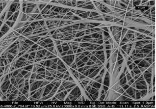
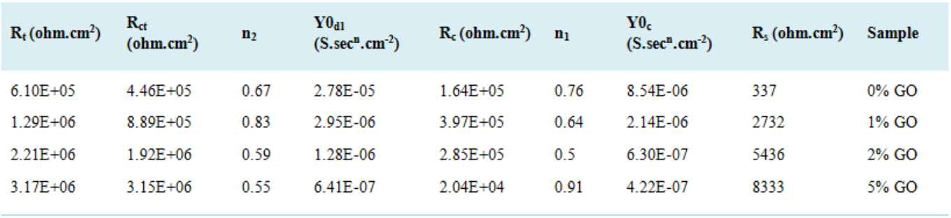
Figure 1. Scanning electron microscope images of the obtained coatings surface at different magnifications.

3.1. Evaluation of Coating Corrosion Resistance

EIS test analysis
The electrochemical impedance test is a test in which alternating current is introduced into the system and the impedance is received from the system in two forms: real and imaginary impedance. By plotting this information in terms of each other in the following different ways, it is possible to analyze the effective parameters in corrosion of the system by fitting the results on the circuit of electrochemical equivalents.
Figure 3 shows the Nyquist curves (Z' in terms of -Z") related to the samples. As shown in Figure 3, in Nyquist curves, the frequency increases from the right side of the graph to the left side of the curve (counter-clockwise), so the point located at the extreme left of the graph has the highest frequency and the highest point. The layer on the right has the lowest frequency . In these curves, the increase in diameter indicates the increase in corrosion resistance of the system, so from the shape of the Nyquist curves, it seems that the sample contains 5% by weight of graphene oxide and then the sample contains 2% by weight of graphene oxide. It has the largest diameter of the semicircle and therefore has the highest resistance to corrosion. Also, the diameter and therefore the resistance of the sample without graphene oxide was lower than the other samples .
In order to better understand the studied system and better check the number of time constants, wind curves should be used. Wind curves for these samples are shown in Figure 4.
In the impedance-module wind curves, the impedance at the lowest frequency can represent the resistance of the entire system against corrosion. Therefore, it is clear from Figure 4 that the impedance at the lowest frequency of the sample containing 5% by weight of graphene oxide is higher than other samples and after that the sample containing 2% by weight of graphene oxide is located. The sample without graphene oxide and the sample containing 1% by weight of graphene oxide have respectively the lowest impedance at the lowest frequency and therefore the lowest total resistance among the investigated samples . The exact value of the resistance of these systems should be determined by modeling these results by the electrochemical equivalent circuit. In order to determine the shape of the electrochemical equivalent circuit, it is necessary to determine the number of time constants of the system. The number of time constants means the number of capacitors/parallel resistors in the electrochemical equivalent circuit. In order to more accurately determine the electrochemical parameters, the results of the impedance test were applied to the electrochemical equivalent circuit in Figure 5.
In the circuit equivalent to 3, there are three resistances (from the left, respectively, solution resistance, coating resistance, and charge transfer resistance) and two fixed phase elements (related to coating and double layer) . In this equivalent circuit, because the plates of the double layer created between the surface of the electrode and the electrolyte are not exactly parallel to each other, and the plate of the electrode and the cover are not completely smooth, a fixed phase element is used instead of an ideal capacitor. The difference between these two elements shows itself in the impedance formula. Capacitor impedance is equal to: Z=1/jωC and this value for CPE is equal to: Z=1/(Y0jω)n.
In these formulas, C means the capacity of the capacitor, ω the phase angle, Y0 the admittance (the opposite of the impedance and equivalent to the capacity parameter in the ideal capacitor) and j is the imaginary term √(-1). As you can see, the difference between the two is only in a power of n, which is a numerical value between zero and one. A value of zero represents an ideal resistance and a value of one represents an ideal capacitor .
Modeling of measured samples with electrochemical equivalent circuit was done by Zsimps software and the results are shown in Figures 1 and 2. As can be seen, the modeling has been able to match the Nyquist and Bud diagrams well, which indicates the reliability of the modeling results.
Considering that the value of the resistance of the whole system (Rt) against corrosion can be obtained from the sum of the coating resistance and the load transfer resistance, it is clear that in the sample containing 5% by weight of graphene oxide, the corrosion resistance is about 3.17 megaohms per square centimeter. which is the highest value of corrosion resistance among the investigated samples. After this sample, the sample containing 2% by weight of graphene oxide with a total resistance of about 2.2 megaohm cm2 has the highest corrosion resistance. The lowest corrosion resistance belongs to the sample without graphene oxide and the sample containing 1% by weight of graphene oxide, which have values of about 0.6 and 1.3 megohm square cm, respectively. Therefore, with the increase in the percentage of graphene oxide in the structure, the corrosion resistance of the system has increased due to the filling of the pores of the coating by these nanoplates .

3.2. Polarization Test Analysis

The polarization test is a useful tool for investigating the thermodynamics and kinetics of corrosion reactions in different systems. In order to more accurately determine the corrosion mechanism in this system, a potentiodynamic polarization test was performed on the investigated samples and is shown in Figure 6. The cathodic (βc) and anodic (βa) branches have been extrapolated and their intersection has given the corrosion current density and corrosion potential (Ecorr). The results are reported in Table 4.
To find the polarization resistance (Rp), Stern-Gray relation is used:
Rp = (βa × βc) / (2.303 × icorr × (βa + βc)) (1)
According to Table 4, the polarization resistance of the sample containing 5% by weight of graphene oxide and the sample containing 2% by weight of graphene oxide is more than the other samples and the sample without graphene oxide is lower than the other samples, which fully confirms the results of the impedance test. In addition, we know that the corrosion potential is related to the thermodynamic tendency to corrosion, and the more negative the potential is, the more active the material becomes and its thermodynamic tendency to corrosion increases . It can be seen here that the thermodynamic tendency to corrosion in samples containing 5% by weight and 2% by weight of graphene oxide is lower than other samples and the thermodynamic tendency to corrosion in samples without graphene oxide is higher than other samples. In other words, increasing the percentage of graphene oxide in the coating has been able to reduce the thermodynamic and kinetic tendency to corrosion in these samples . This improvement in the corrosion properties of the coating with an increase in the percentage of graphene oxide is due to the improvement of the barrier properties of the coating due to the placement of graphene oxide nano sheets in the pores of the coating, which prevented the penetration of the electrolyte into the coating and increased its corrosion resistance .
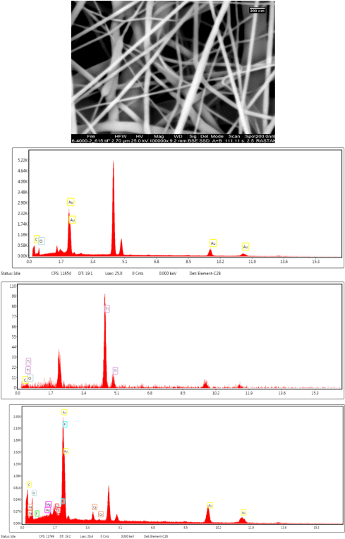
Figure 2. EDX elemental analysis image of polycaprolactone-graphene oxide composite.
Figure 3. The Nyquist curves of the sample and the fit results of the data obtained from the EIS test on the appropriate electrochemical equivalent circuit related to the examined samples (the points are the results of the test and the lines are the results of the fit).
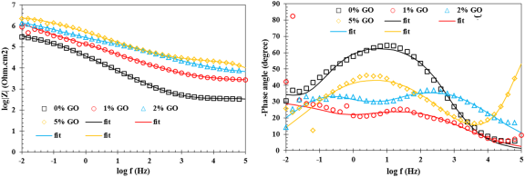
Figure 4. Curves of (a) wind-impedance modulus and (b) wind-phase angle of the samples related to the examined samples (the points are the results of the test and the lines are the results of the fit).
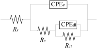
Figure 5. Electrochemical equivalent circuit used to model the impedance test of the investigated samples.
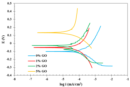
Figure 6. The results of the potentiodynamic polarization test of the examined samples to extract the corrosion current density value (icorr), Tafeli area.
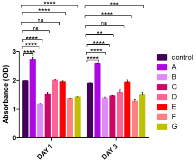
Figure 7. MTT test results in two intervals of 24 and 72 hours.

3.3. Assessment of Cytotoxicity Behavior by MTT-Assay

With the progress of biological sciences, measuring the rate of proliferation, survival and death of cells under different conditions has become very important. In this regard, MTT analysis has made a great contribution to the study of biocompatibility of various materials by providing a non-radioactive and colorimetric system with high safety .
Cytotoxicity tests are tests that check the unwanted effects of various compounds on cells. These processes are carried out in the environment outside the human body or so-called extracorporeal. Most of these processes also use cell culture.
The MTT test is the most well-known test to check cell viability. The main purpose of this test is to investigate the toxicity effect of compounds, drugs or other supplements on the cell. Of course, it may also be mentioned in the articles as a process for checking cell proliferation or counting.
MTT analysis is able to distinguish between living and dead cells by affecting intracellular organelles. In this method, after being cultivated in the laboratory, the cells are "treated" with the desired substances in order to evaluate their toxicity .
At the end of this test, for each concentration of the substance, the cell viability is determined. Although this method is originally for solutions and compounds soluble in water, it is currently implemented for other compounds soluble in organic solvents and nanoparticles .
In the MTT test, which used the indirect MTT method, we divided it into two periods of 24 and 72 hours. Once in 24 hours, we performed the MTT method for 3 composite compositions including polycaprolactone/graphene oxide 1%, polycaprolactone/graphene oxide 2% and polycaprolactone/graphene oxide 5%, and the following results were obtained.
Group D, which included 2% polycaprolactone/graphene oxide, did not cause significant cell death on the first day.
But groups C and D showed toxicity on the first day (24 hours) and also on the third day (72 hours).
The same group D showed significant lethality on the third day (72 hours).
In the analysis of graphs, the results of the MTT test indicated the toxicity of C and F scaffolds in the period of one and three days after the treatment compared to the control condition (TCP). (*p<0.05, **p<0.01, ***p<0.001, ****p<0.0001). Group D did not have significant lethality on the cells on day 1, but it caused significant lethality on day 3.
4. Conclusion
By creating a polycaprolactone-graphene oxide nanofiber composite coating, the biological and antibacterial properties of titanium alloy will be improved and its corrosion resistance will probably change. In this project, the main question is extracting effective parameters in creating a composite coating on titanium surface by electrospinning method and characterizing and biological evaluation of the created coating.
Abbreviations
PCL: Polycaprolactone
GO: Graphene Oxide
Acknowledgments
In this article, we thank Mr. Alireza Khalili for her efforts to edit the article.
Declaration
Ethics approval and consent to participate. All procedures performed in studies involving human participants were in accordance with the ethical standards of the institutional and national research committee. This article does not contain any studies with animals performed by any of the authors.
Conflicts of Interest
The authors declare no conflict of interest.
References
[1] J. B. Park, J. D. Bronzino, "Biomaterials, Principles and Applications", CRC Press, 2002, 21.
[2] A. Rabeie, B. Thomas, B. Neville, J. W. Lee, J. W Coumo, " Microstructure, Mechanical Properties and Biological Responses to Functionally Graded HA Coatings", Materials Science and Engineering C, 27(2007) 523.
[3] H. R. Bakhsheshi-Rad, Z. Hadisi, E. Hamzah, A. F. Ismail, M. Aziz, M. Kashefian, Drug delivery and cytocompatibility of ciprofloxacin loaded gelatin nanofibers-coated Mg alloy, Materials Letters, 2017.
[4] Wang, X., Ding, B. and Li, B., “Biomimetic electrospun nanofibrous structures for tissue engineering,” Materials Today, Vol. 16, pp. 229–241, 2013.
[5] Woodruff, M. A., and Hutmacher, W. H., “The return of a forgotten polymer : Polycaprolactone in the 21st century”, Progress in Polymer Science, Vol. 35, pp. 1217–1256, 2010.
[6] Li M, Wang Y, Liu Q, Li Q, Cheng Y, Zheng Y, et al. In situ synthesis and biocompatibility of nano hydroxyapatite on pristine and chitosan functionalized graphene oxide. J Mater Chem B 2013; 1(4): 475–84.
[7] Neelgund GM, Oki A, Luo Z. In-situ deposition of hydroxyapatite on graphene nanosheets. Mater Res Bull 2013; 48(2): 175–9.
[8] Rodrı´guez-Lorenzo Luis M, Benito-Garzo´n Lorena, Barroso- Bujans Fabienne, Ferna´ndez Mar. Synthesis and biocompatibility of hydroxyapatite in a graphite oxide.
[9] Ramakrishna, S., Fujihara, K. Teo, W. Yong, T. and Ramaseshan, R. “Electro spunnan of ibers : solving global issues,” Materials today, Vol. 9, pp. 40–50, 2006.
[10] Hutmacher D. W. and Dalton P. D., Melt Electrospinning, Chem. Asian J., 6, 44-56, 2011.
[11] Lee S. and Obendorf S. K., Developing Protective Textile Materials as Barriers to Liquid Penetration Using Melt-Electrospinning, J. Appl. Polym. Sci., 102, 3430-3437, 2006.
[12] Ko J., Kan D., and Jun M. B., Combining Melt Electrospinning and particulated leaching for fabrication of porous microfibers, Manuf. Lett., 3, 5-8, 2015.
[13] Karchin A., Simonovsky F. I., Ratner B. D., and Sanders J. E., Melt Electrospinning of Biodegradable Polyurethane Scaffolds, Acta Biomater., 7, 3277-3284, 2011.
[14] Hochleitner G., Jungst T., Brown T. D., Hahn K., Moseke C., Jakob F., Dalton P. D., and Groll J., Additive Manufacturing of Scaffolds with Sub-Micron Filaments via Melt Electrospinning Writing, Biofabrication, 7, 35002, 2015.
[15] Lyons J., Li C., and Ko F., Melt-Electrospinning Part I: Processing Parameters and Geometric Properties, Polymer, 45, 7597-7603, 2004.
[16] Hochleitner G., Hummer J. F., Luxenhofer R., and Groll J., high definition fibrous poly(2-Ethyl-2-oxazoline) scaffolds through melt electrospinning writing, Polymer, 55, 5017-5023, 2014.
[17] Dalton P. D., Grafahrend D., Klinkhammer K., Klee D., and Moller M., Electrospinning of Polymer Melts: Phenomenological Observations, Polymer, 48, 6823-6833, 2007.
[18] Lyons J. and Ko F., Melt Electrospinning of Polymers: A Review, Polym. News, 30, 170-178, 2005.
[19] Kong C. S., Jo K. J., Jo N. K., and Kim H. S., Effects of The Spin line temperature profile and melt index of poly on Melt-Electrospinning, Technical Report, 2009.
[20] Reneker D., Kataphinana W., Theron A., Zussman E., nanofiber garlands of polycaprolactone by electrospinning, Polymer, 43, 6785-6794, 2002.
[21] Li X., Liu H., Wang J., and Li C., Preparation and Characterization of polycaprolactone nonwoven mats via melt electrospinning, Polymer, 53, 248-253, 2012.
[22] Ma M., Mao Y., Gupta M., Gleason K. K., and Rutledge G. C., Superhydrophobic Fabrics Produced by Electrospinning and Chemical Vapor Deposition, Macromolecules, 38, 9742-9748, 2005.
[23] Muerza-Cascante M. L., Haylock D., Hutmacher D. W., and Dalton P. D., Melt Electrospinning and Its Technologization in Tissue Engineering, Tissue Eng., Part B: Rev., 21, 187-202, 2014.
[24] Detta N. et al., Melt Electrospinning of Polycaprolactone and Its Blends with Poly (ethylene glycol), Polym. Int., 59, 1558- 1562, 2010.
[25] Li X., Wang Z., Wang J., Liu J., and Li C., Preparation and properties of TPU micro/nanofibers by A layser melt-Elec trospinning System, Polym. Eng. Sci., 54, 1412-1417, 2014.
[26] Lyons J. M., Melt-Electrospinning of Thermoplastic Polymers: An Experimental and Theoretical Analysis, Ph. D. Thesis, Drexel University, October 2004.
[27] Bhardwaj N. and Kundu S. C., Electrospinning: A Fascinating Fiber Fabrication Technique, Biotech. Adv., 28, 325-347, 2010.
[28] Li X., Zhang Y., Li H., Chen H., Ding Y., and Yang W., Effect of Oriented Fiber Membrane Fabricated via Needleless Melt electrospinning on water filtration efficiency Desalination, 344, 266-273, 2014.
[29] Zhou H., Green T. B., and Joo Y. L., The Thermal Effects on Electrospinning of Polylactic Acid Melts, Polymer, 47, 7497-7505, 2006.
[30] McCann J. T., Marquez M., and Xia Y., Melt Coaxial Electrospinning: A Versatile Method for The Encapsulation of Solid materials and fabrication of phase change nanofibers, Nano lett., 6, 2868-2872, 2006.
[31] Dalton P. D., Joergensen N. T., Groll J., and Moeller M., Patterned Melt Electrospun Substrates for Tissue Engineering, Biomed. Mater., 3, 1-11, 2008.
[32] Zong H., Xia X., Liang Y., Dai S., Alsaedi A., Hayat T., Kong F., and Hong Pan J., Designing Function-Oriented Artificial Nanomaterials and Membranes via Electrospinning and Electrospraying Techniques, Mater. Sci. Eng., C, 2017.
[33] Li X., Liu H., Wang J., and Li C., Preparation and Characterization of PLLA/nHA Nonwoven Mats via Laser Melt Electrospinning, Mater. Lett., 73, 103-106, 2012.
[34] Dutta P. K., Dutta J., and Tripathi V., Chitin and Chitosan: Chemistry, Properties and Applications, j. Sci. Ind. Res., 63, 20-31, 2004.
[35] Sill T. J. and von Recum H. A., Electrospinning: Applications in Drug Delivery and Tissue Engineering, Biomaterials, 29, 1989-2006, 2008.
[36] Ko J., Khadem Mohtaram N., Ahmed F., Montgomery A., Carlson M., Lee P. C. D., Willerth S. M., and Jun M. B. G., Fabrication of poly caprolactone microfiber scaffolds with Varying Topography and Mechanical Properties for Stem Cell-Based Tissue Engineering Applications, J. Biomater. Sci., Polym. Ed., 25, 1-17, 2014.
[37] Cho D., Zhou H., Cho Y., Audus D., and Joo Y. L., Structural Properties and Superhydrophobicity of Electrospun Polypropylene Fibers from Solution and Melt, Polymer, 51, 6005- 6012, 2010.
[38] Ma M., Hill R. M., Lowery J. L., Fridrikh S. V., and Rutledge G. C., Electrospun Poly(Styrene-Block-Dimethylsiloxane) Block Copolymer Fibers Exhibiting Superhydrophobicity, Langmuir, 21, 5549-5554, 2005.
[39] singh., steely l., and ALLcock H. R., poly [bis(2,2,2-trifluo-roethoxy)phosphaz] superhydrophobic Nanofibers Langmuir, 21, 11604-11607, 2005.
[40] Iqbal-Sabir M., Xu X., and Li L., A Review on Biodegradable Polymeric Materials for Bone Tissue Engineering Applications, J. Mater. Sci., 44, 5713-5724, 2009.
Cite This Article
  • APA Style

    Fazlali, P., Tahernejad, M., Biglari, L., Eslami, M. (2024). Surface Modification of Ti-6Al-4V Alloy by Polycaprolactone-Graphene Oxide Composite Coating . International Journal of Materials Science and Applications, 13(2), 13-23. https://doi.org/10.11648/j.ijmsa.20241302.11

    Copy | Download

    ACS Style

    Fazlali, P.; Tahernejad, M.; Biglari, L.; Eslami, M. Surface Modification of Ti-6Al-4V Alloy by Polycaprolactone-Graphene Oxide Composite Coating . Int. J. Mater. Sci. Appl. 2024, 13(2), 13-23. doi: 10.11648/j.ijmsa.20241302.11

    Copy | Download

    AMA Style

    Fazlali P, Tahernejad M, Biglari L, Eslami M. Surface Modification of Ti-6Al-4V Alloy by Polycaprolactone-Graphene Oxide Composite Coating . Int J Mater Sci Appl. 2024;13(2):13-23. doi: 10.11648/j.ijmsa.20241302.11

    Copy | Download

  • @article{10.11648/j.ijmsa.20241302.11,
      author = {Paria Fazlali and Mahrokh Tahernejad and Leila Biglari and Mahla Eslami},
      title = {Surface Modification of Ti-6Al-4V Alloy by Polycaprolactone-Graphene Oxide Composite Coating
    },
      journal = {International Journal of Materials Science and Applications},
      volume = {13},
      number = {2},
      pages = {13-23},
      doi = {10.11648/j.ijmsa.20241302.11},
      url = {https://doi.org/10.11648/j.ijmsa.20241302.11},
      eprint = {https://article.sciencepublishinggroup.com/pdf/10.11648.j.ijmsa.20241302.11},
      abstract = {In this research, Polycaprolactone-graphene oxide Nanocomposite coating was synthesized and characterized by Electrospinning on Ti6AL4V alloy. In order to create a uniform coating with optimal thickness, the effective parameters of Electrospinning coating, including solvent, polymer concentration, and bioceramic percentage, were investigated. Also, the cytotoxicity and corrosion tests were evaluated by the electrochemical polarization test method of the created coating in comparisons and different percentages. In order to characterize the coating, a test such as a scanning electron microscope was used. The results showed that as much as the amount of Graphene oxide is increased, the diameter of Nanofibers decreases. The diameter of Polycaprolactone Nanofibers was 1.3 micrometers, which increases to 56.0 micrometers by adding Graphene oxide. The results of the corrosion test showed that the use of Nano composite coating increased the corrosion resistance to the size of the coating. The nanocomposite coating consists of polycaprolactone nanofibers and graphene oxide, which mimics the behavior of the extracellular matrix and improves the biological and antibacterial behavior of the titanium surface. So far, there has been no report on the creation of this fibrous nanocomposite coating on titanium. The results of the cytotoxicity test showed that the use of Nanocomposite coating has effectively reduced the cytotoxicity on the scaffolds. By creating a polycaprolactone-graphene oxide nanofiber composite coating, the biological and antibacterial properties of titanium alloy will be improved and its corrosion resistance will probably change. In this project, the main question is extracting effective parameters in creating a composite coating on titanium surface by electrospinning method and characterizing and biological evaluation of the created coating.
    },
     year = {2024}
    }
    

    Copy | Download

  • TY  - JOUR
    T1  - Surface Modification of Ti-6Al-4V Alloy by Polycaprolactone-Graphene Oxide Composite Coating
    
    AU  - Paria Fazlali
    AU  - Mahrokh Tahernejad
    AU  - Leila Biglari
    AU  - Mahla Eslami
    Y1  - 2024/04/02
    PY  - 2024
    N1  - https://doi.org/10.11648/j.ijmsa.20241302.11
    DO  - 10.11648/j.ijmsa.20241302.11
    T2  - International Journal of Materials Science and Applications
    JF  - International Journal of Materials Science and Applications
    JO  - International Journal of Materials Science and Applications
    SP  - 13
    EP  - 23
    PB  - Science Publishing Group
    SN  - 2327-2643
    UR  - https://doi.org/10.11648/j.ijmsa.20241302.11
    AB  - In this research, Polycaprolactone-graphene oxide Nanocomposite coating was synthesized and characterized by Electrospinning on Ti6AL4V alloy. In order to create a uniform coating with optimal thickness, the effective parameters of Electrospinning coating, including solvent, polymer concentration, and bioceramic percentage, were investigated. Also, the cytotoxicity and corrosion tests were evaluated by the electrochemical polarization test method of the created coating in comparisons and different percentages. In order to characterize the coating, a test such as a scanning electron microscope was used. The results showed that as much as the amount of Graphene oxide is increased, the diameter of Nanofibers decreases. The diameter of Polycaprolactone Nanofibers was 1.3 micrometers, which increases to 56.0 micrometers by adding Graphene oxide. The results of the corrosion test showed that the use of Nano composite coating increased the corrosion resistance to the size of the coating. The nanocomposite coating consists of polycaprolactone nanofibers and graphene oxide, which mimics the behavior of the extracellular matrix and improves the biological and antibacterial behavior of the titanium surface. So far, there has been no report on the creation of this fibrous nanocomposite coating on titanium. The results of the cytotoxicity test showed that the use of Nanocomposite coating has effectively reduced the cytotoxicity on the scaffolds. By creating a polycaprolactone-graphene oxide nanofiber composite coating, the biological and antibacterial properties of titanium alloy will be improved and its corrosion resistance will probably change. In this project, the main question is extracting effective parameters in creating a composite coating on titanium surface by electrospinning method and characterizing and biological evaluation of the created coating.
    
    VL  - 13
    IS  - 2
    ER  - 

    Copy | Download

Author Information
  • Department of Biomedical Engineering, Medical Tehran Branch, Islamic Azad University, Teran, Iran

  • Department of Biomedical Engineering, Research and Science Branch, Islamic Azad University, Tehran, Iran

  • Department of Biomedical Engineering, University of Shahrekord, Shahrekord, Iran

  • Department of Biomedical Engineering, Nooshirvani University, Mazandaran, Iran