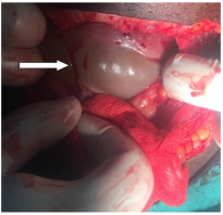
Background: We are presenting a 27 years old female with her second pregnancy at 42 weeks of gestation who was admitted because she was post date with one previous uterine scar and planned for emergency caesarean section due to aforementioned reasons. She was prepared and sent to theatre where uterine scar dehiscence was found but she got a live fetus male baby weighed 3.2kg and scored 9 and 10 at 1st and 5th minutes respectively. The patient received all necessary post operative cares, she progress well while in the ward and was discharged in good health after 3 days and came again in the 7th day for suture remove and then continued with post natal visits as per protocol until was discharged from the clinic. Conclusion: Uterine scar dehiscence without notable complications to the mother and her fetus is rare condition which necessitates serious attention to most women with previous caesarean delivery. In preconception period around 3 to 6 months post previous c/section, transvaginal ultrasound is ideal to measure the lower uterine segment thickness and during pregnancy is better at 32 to 36 weeks using trans-abdominal ultrasound. Whenever an ultrasound is not conclusive, MRI can be used. This case is presented to emphasize on importance of scheduled caesarean section at 37 completed weeks.
| Published in | Journal of Gynecology and Obstetrics (Volume 14, Issue 3) |
| DOI | 10.11648/j.jgo.20261403.11 |
| Page(s) | 71-74 |
| Creative Commons |
This is an Open Access article, distributed under the terms of the Creative Commons Attribution 4.0 International License (http://creativecommons.org/licenses/by/4.0/), which permits unrestricted use, distribution and reproduction in any medium or format, provided the original work is properly cited. |
| Copyright |
Copyright © The Author(s), 2026. Published by Science Publishing Group |
Caesarean Section, Uterine Scar Dehiscence, Live Fetus
C/S | Caesarean Section |
HIV | Human Immunodeficiency Virus |
Kg | Kilogram |
MRI | Magnetic Resonance Imaging |
VDRL | Venerial Disease Research Laboratory |
WHO | World Health Organisation |
| [1] | Simran A et al. Examining the role and relevance of the critical analysis and comparison of cesarean section rates in a changing world. |
| [2] | Saifon Chawanpaiboon et al. Severe Complications of Uterine Dehiscence Post-Lower Segment Cesarean Section: A Case Report Emphasizing the Importance of Timely Diagnosis and Intervention. |
| [3] | Bashiri A, Burstein E, Rosen S, et al. Clinical significance of uterine scar dehiscence in women with previous cesarean delivery: Prevalence and independent risk factors. |
| [4] | Mohamad K. Ramadana et at. Incidence and Risk Factors of Uterine Scar Dehiscence Identified at Elective Repeat Cesarean Delivery: A Case-Control Study. J Clin Gynecol Obstet. 2018; 7(2): 37-42. |
| [5] | Zeb L. Frequency of scar dehiscence in patients with previous one caesarean section having scar tenderness. J Khyber Coll Dentist. 2023; 13(3): 45-8. |
| [6] | Tyagi N, Prabhakar M, Tyagi S. Retrospective study to find predictive factors of scar dehiscence in previous caesarean section to prevent maternal and perinatal morbidity and mortality. Int J Reproduct Contracept Obstetr Gynecol. 2019; 8(2): 531-6. |
| [7] | Kaplanoglu M, Bulbul M, Kaplanoglu D, Bakacak SM. Effect of multiple repeat cesarean sections on maternal morbidity: data from southeast Turkey. I nt MedJ Experiment Clin Res. 2015; 21: 1447. |
| [8] | Chen SH, Du XP. Silent spontaneous posterior uterine rupture of a prior caesarean delivery at 36weeks of gestation. BMC Pregnancy Childbirth. 2019; 19(1): 4–6. |
| [9] | Hamar BD, Levine D, Katz NL, Lim KH. Expectant management of uterine dehiscence in the second trimester of pregnancy. Obstet Gynecol. 2003; 102 (5SUPPL.): 1139–42. |
| [10] | Fox NS, Gerber RS, Mourad M, Saltzman DH, Klauser CK, Gupta S, et al. Pregnancy outcomes in patients with prior uterine rupture or dehiscence. ObstetGynecol. 2014; 123(4): 785–9. |
| [11] | Hofmeyr GJ, Say L, Gülmezoglu AM. WHO systematic review of maternal mortality and morbidity: The prevalence of uterine rupture. BJOG An IntJ Obstet Gynaecol. 2005; 112(9): 1221–8. |
| [12] | Mrema S, Massinde A, Matovelo D, Kihunrwa A, Rumanyika R, Kidenya B. Fetomaternal outcomes in patients with Uterine Rupture managed at Bugando Medical Centre, Tanzania: 5-year review of cases. 2019; 1–23. |
| [13] | Sawada M, Matsuzaki S, Nakae R, Iwamiya T, Kakigano A, Kumasawa K, et al. Treatment and repair of uterine scar dehiscence during cesarean section. Clin Case Reports. 2017; 5(2): 145–9. |
| [14] | Chapman SJ, Owen J, Hauth JC. One-versus two-layer closure of a low transverse cesarean: The next pregnancy. Obstet Gynecol. 1997; 89(1): 16–8. |
| [15] | Klemm P, Koehler C, Mangler M, Schneider U, Schneider A. Laparoscopic and vaginal repair of uterine scar dehiscence following cesarean section as detected by ultrasound. J PerinatMed. 2005; 33(4): 324–31. |
| [16] | Bromley B, Pitcher BL, Klapholz H, Lichter E, Benacerraf BR. Sonographic appearance of uterine scar dehiscence. IntJ Gynecol Obstet. 1995; 51(1): 53–6. |
| [17] | Ishan Kumar, Ashish Verma et al. Utility of multiparametric MRI in c/section scar characterization and preoperative prediction of scar dehiscence: a prospective study. |
APA Style
Mvungi, E. D., Kaiza, I. L., Mabega, N. G. (2026). Uterine Scar Dehiscence Found During Emergency Caesarean Section: A Case Report. Journal of Gynecology and Obstetrics, 14(3), 71-74. https://doi.org/10.11648/j.jgo.20261403.11
ACS Style
Mvungi, E. D.; Kaiza, I. L.; Mabega, N. G. Uterine Scar Dehiscence Found During Emergency Caesarean Section: A Case Report. J. Gynecol. Obstet. 2026, 14(3), 71-74. doi: 10.11648/j.jgo.20261403.11
@article{10.11648/j.jgo.20261403.11,
  author = {Emiliana Dismas Mvungi and Innocent Lutakyamilwa Kaiza and Ndakibae Gabriel Mabega},
  title = {Uterine Scar Dehiscence Found During Emergency Caesarean Section: A Case Report},
  journal = {Journal of Gynecology and Obstetrics},
  volume = {14},
  number = {3},
  pages = {71-74},
  doi = {10.11648/j.jgo.20261403.11},
  url = {https://doi.org/10.11648/j.jgo.20261403.11},
  eprint = {https://article.sciencepublishinggroup.com/pdf/10.11648.j.jgo.20261403.11},
  abstract = {Background: We are presenting a 27 years old female with her second pregnancy at 42 weeks of gestation who was admitted because she was post date with one previous uterine scar and planned for emergency caesarean section due to aforementioned reasons. She was prepared and sent to theatre where uterine scar dehiscence was found but she got a live fetus male baby weighed 3.2kg and scored 9 and 10 at 1st and 5th minutes respectively. The patient received all necessary post operative cares, she progress well while in the ward and was discharged in good health after 3 days and came again in the 7th day for suture remove and then continued with post natal visits as per protocol until was discharged from the clinic. Conclusion: Uterine scar dehiscence without notable complications to the mother and her fetus is rare condition which necessitates serious attention to most women with previous caesarean delivery. In preconception period around 3 to 6 months post previous c/section, transvaginal ultrasound is ideal to measure the lower uterine segment thickness and during pregnancy is better at 32 to 36 weeks using trans-abdominal ultrasound. Whenever an ultrasound is not conclusive, MRI can be used. This case is presented to emphasize on importance of scheduled caesarean section at 37 completed weeks.},
 year = {2026}
}
TY  - JOUR
T1  - Uterine Scar Dehiscence Found During Emergency Caesarean Section: A Case Report
AU  - Emiliana Dismas Mvungi
AU  - Innocent Lutakyamilwa Kaiza
AU  - Ndakibae Gabriel Mabega
Y1  - 2026/06/12
PY  - 2026
N1  - https://doi.org/10.11648/j.jgo.20261403.11
DO  - 10.11648/j.jgo.20261403.11
T2  - Journal of Gynecology and Obstetrics
JF  - Journal of Gynecology and Obstetrics
JO  - Journal of Gynecology and Obstetrics
SP  - 71
EP  - 74
PB  - Science Publishing Group
SN  - 2376-7820
UR  - https://doi.org/10.11648/j.jgo.20261403.11
AB  - Background: We are presenting a 27 years old female with her second pregnancy at 42 weeks of gestation who was admitted because she was post date with one previous uterine scar and planned for emergency caesarean section due to aforementioned reasons. She was prepared and sent to theatre where uterine scar dehiscence was found but she got a live fetus male baby weighed 3.2kg and scored 9 and 10 at 1st and 5th minutes respectively. The patient received all necessary post operative cares, she progress well while in the ward and was discharged in good health after 3 days and came again in the 7th day for suture remove and then continued with post natal visits as per protocol until was discharged from the clinic. Conclusion: Uterine scar dehiscence without notable complications to the mother and her fetus is rare condition which necessitates serious attention to most women with previous caesarean delivery. In preconception period around 3 to 6 months post previous c/section, transvaginal ultrasound is ideal to measure the lower uterine segment thickness and during pregnancy is better at 32 to 36 weeks using trans-abdominal ultrasound. Whenever an ultrasound is not conclusive, MRI can be used. This case is presented to emphasize on importance of scheduled caesarean section at 37 completed weeks.
VL  - 14
IS  - 3
ER  -
Department of Obstetrics and Gynecology, Sekou Toure Regional Referral Hospital, Mwanza, United Republic of Tanzania
Department of Clinical Research, National Institute for Medical Research, Mwanza, United Republic of Tanzania
Information