Research Article | | Peer-Reviewed

Two News Species of Psyllids Genus Syntomoza (Hemiptera: Liviidae) Pest Insects Associated with Flacourtia Indica (Flacourtiaceae) in Cameroon

Received: 25 December 2025     Accepted: 8 January 2026     Published: 4 February 2026
Views:       Downloads:
Abstract

Background: Psyllids mainly through their larvae which suck sap from their host plants can be harmful by causing the transmission of diseases through their saliva. Objective: The aim of this study was to describe the news species of psyllids genus Syntomoza pest insects associated with Flacourtia indica (Flacourtiaceae). Methods: Psyllids specimens were collected using an entomological net and a mouth aspirator. The specimens were preserved in 70% ethanol, mounted on slides. Identifications, measurements and illustrations were made using a Microscope. Results: Syntomoza biniae sp. nov., have vertex sub-rectangular without foveae; male proctiger in profile, with lateral lobe large, broadly rounded in the middle, narrowed apically, obliquely truncate at apex; paramere with apical region narrow curved forward bearing two short black thick spine oriented inwards; inner face with a conspicuous fold anteriorly extending obliquely toward apex; distal segment of aedeagus almost curved in basal half, dorsal margin weakly sinuate; female subgenital plate in lateral view subtriangular, very enlarged at the base with distinct hump; dorsal and ventral valvulae slightly curved dorsally. Fifth instar larvae, with antennal segments with following numbers of pointed setae: 1(0), 2(0), 3(2), 4(0), 5(1), 6(0), 7(2); dorsum of caudal plate bearing scarcely arranged small sharp cuticular teeth; anus ventral, surrounded by only outer circumanal pore ring. Syntomoza flacourtiae sp. nov., with male proctiger in profile medially with well-developed, broadly rounded posterior expansions, rather with two pits, one at base and other at apex, narrowed apically, apical margin rounded; paramere is rather with bulging posterior margin on one-third basally, anterior margin incurved, inner face covered with two row of eight stouter setae situated along anterior margin medially; circumanal ring oval to pear shaped. In fifth instar larvae: antenna segments 3 and 5 without numbers of pointed setae; forewing pad with a row of seven marginal long lanceolate; abdomen on all dorsum surface bearing lanceolate setae and additional like-scaly setae; anus terminal, without additional pore fields. Damage caused by Syntomoza spp. includes distortion and winding of the lateral edges of young leaves. Conclusion: Taxonomic studies indicated that, Syntomoza biniae sp. nov. and Syntomoza flacourtiae sp. nov. are the news species of Liviidae family described from Adamawa Region of Cameroon.

Published in American Journal of Zoology (Volume 9, Issue 1)
DOI 10.11648/j.ajz.20260901.12
Page(s) 8-22
Creative Commons

This is an Open Access article, distributed under the terms of the Creative Commons Attribution 4.0 International License (http://creativecommons.org/licenses/by/4.0/), which permits unrestricted use, distribution and reproduction in any medium or format, provided the original work is properly cited.

Copyright

Copyright © The Author(s), 2026. Published by Science Publishing Group

Keywords

Syntomoza Biniae sp. nov., Syntomoza Flacourtiae sp. nov., Psyllid Pest, Flacourtia Indica, Flacourtiaceae, Adamawa-Cameroon

1. Introduction
Psyllids (Hemiptera: Psylloidea) are phloem-feeding insects, they are generally highly host specific on woody dicotyledonous plants and display their largest species diversity in the tropics . Most of the known psyllids species in the world are oligophagous being restricted to one or a few closely related hosts plants, particularly in their immature stages . Psyllids can be harmful to their plant hosts mainly through their larvae which take sap mainly from the flower buds causing the transmission of diseases through their saliva . The intake of food leads to the alteration of photosynthetic activity, with consequent disturbed growth and the weakening of the plant which becomes sensitive to attacks by secondary organisms .
Syntomoza Crawford genus belonging to the Liviinae (Paurocephalinae) subfamily and to Liviidae family . The Liviidae family included Euphylleurinae Crawford, 1914, Liviinae Löw, 1879 and Neophyllurinae subfam. nov. The Liviinae subfamily included six genera: Aphorma Hodkinson, Camarotoscena Haupt, Diclidophlebia Crawford, Livia Latreille, Paurocephala Crawford and Syntomoza Enderlein. Keys for the identification of adults and fifth instar larvae are provided by Burckhardt & Mifsud . From Cameroon, some species belonging of Liviidae family have been described: Diclidophlebia leptonychiae and D. irvingiae ; D. eastopi xuani ; D. xuani Messi et al. ; D. andjigae ; Paurocephala sinuate and P. nkomekui and P. famendongoeis .
Seven species belonging to Syntomoza genus have been described worldwide, one of them in Africa, Syntomoza africana collected on Dovyalis abyssinica (Flacourtiaceae) in Ethiopia, Kenya and Uganda . The host plants of Syntomoza species belonging to the Flacourtiaceae family, except for S. unicolor which is associated with Populus spp. belonging to Saliaceae family.
The knowledge on the diversity of psyllids fauna and their distribution in Cameroon is still incomplete and it general poorly investigated. During surveys in the Adamawa region of Cameroon, two psyllids species of Syntomoza genus was collected on Flacourtia indica Willd. (Flacourtiaceae); this plant family is also quite represented in the Adamawa region of Cameroon. This paper report for the first time from Cameroon, on the description of the news species of psyllids genus Syntomoza (Hemiptera: Liviidae) pest insects associated with Flacourtia indica (Flacourtiaceae).
2. Materials and Methods
Type locality: University Campus of Ngaoundere, 7°25’09, 8’’N; 13°32’42, 4’’E; 1085 m. Type material: Holotype: ♂, University Campus of Ngaoundere, 28 June 2011. Paratype: Beka-Hossere: 2♂, 4♀, 7°20’19, 9’’N; 13°33’31, 4’’E; 1111 m, 10 July 2015. Bini: 16♂, 16♀, 1larva, 14-24 May 2011; 51♂, 42♀, 1larva, 2-11-23 June 2011; 19♂, 11♀, 12-21-30 July 2011; 1♀, 2 January 2012; 3♂, 5♀, 23 March 2012; 2♂, 3♀, 1 larva, 3 April 2012; 6♂, 8♀, 16-25 August 2012; 1♂, 1♀, 3 September 2012; 4♂, 7♀, 29 March 2013; 9♂, 4♀, 3larvae, 3 April 2013; 10♂, 7♀, 26 July 2015. University Campus of Ngaoundere: 26♂, 20♀, 21-31 May 2011; 32♂, 22♀, 9-28 June 2011; 21♂, 16♀, 7-24 July 2011; 48♂, 24♀, 4-13-20 August 2011; 6♂, 2♀, 14-23 August 2012; 3♀, 26 December 2012; 4 larvae, 14 September 2024. Dang: 15♂, 12♀, 1larva, 17-26 v 2011; 31♂, 41♀, 1larva, 4-14-25 vi 2011; 7♂, 7♀, 5 July 2011; 3♂, 4♀, 2-11 August 2011; 4♂, 5♀, 4larvae, 31 March 2012; 12♂, 20♀, 4larvae, 5 April 2012; 1♂, 2♀, 11-30 August 2012; 2 larvae, 30 March 2013; 8♂, 10♀, 1 larva, 4 April 2013, 7°24’42, 4’’N; 13°32’42, 4’’E; 1077 m.
Type series deposit: The type series of Syntomoza spp. (Figures 1a-d) were deposited in the collections of the Laboratory of Zoology, Higher Teachers’ Training College, University of Yaounde I, Cameroon (LZUY).
Field survey: The observations and survey took place in various localities of the Adamawa Region, Cameroon, from May 2011 to September 2024. During each survey, host plants were inspected. Adults of psyllids were captured with a mouth aspirator. Nymphs were sampled directly from buds and leaves of the host plant. The host plant was identified at the National Herbarium in Yaounde (Cameroon) and is deposited in LZUY.
Observations and illustrations: The specimens are preserved dry and slide-mounted or in 70% ethanol and are deposited in Laboratory of Zoology, University of Yaounde I. The morphology was illustrated using transmission Leica microscope and measurements were made from slide-mounted using Leica stereomicroscope.
Terminologies: The terminologies used for the description follow the identification keys of Mifsud & Burckhardt and Burckhardt & Mifsud .
Head: Vertex, median suture, genal cones, ocellus, compound eyes; Antenna: Rhinaria, flagellomere; Wing: costal break, pterostigma, vein, spinules, cells; Leg: metacoxa, meracanthus, metatibia, spurs, arolium; Male genitalia: proctiger, paramere, aedeagus, ductus ejaculatorius; Female genitalia: proctiger, subgenital plate, dorsal and ventral valvulae, circumanal ring.
3. Results
3.1. Description of Syntomoza Biniae sp. nov.
Adult Coloration: (Figures 1a and 1b), head and the thorax light brown; the rest of the body dark yellow; the last two segments of the antenna are dark. The compound eyes are dark red.
Structure: Head (Figure 2) less broad than body width, slightly less than body width 0.8-1.12 mm in males and 0.8-1.0 mm in females. Head is curved perpendicularly with respect to the longitudinal axis of the body; the vertex is almost rectangular, flat, uniformly rounded forward and downward, it bears small dense simple setae in its middle on either side of the median suture; genal cones conspicuously separated from vertex, emerging in same plane without basal suture, very short and anteriorly broadly rounded, divergent apically outwards, obtusely rounded at apex; the anterior margin each bearing a long simple setae. The posterior ocelli at the extreme lateral base of vertex; anterior ocellus larger than posterior pair, visible in front; eyes moderately large, hemispherical and somewhat recessive.
Antenna (Figure 4) is longer than the width of the head; ten segmented, basal two segments robust, cylindrical, the third segment is the longest; fourth to eighth segments are subequal to each other, the two terminal segments equal in length but wider than the preceding, terminal segment with two almost equal long simple setae at apex, two small simple setae present near each rhinaria on segment 4, 6 and 9.
Thorax dorsally convex and curving forward into head, without pubescence, pronotum flat, viewed dorsally, longer in middle and narrowed laterally, convexly rounded.
Forewings (Figure 6) is membranous and covered with dense spinule surfaces occupying all the surfaces of the wing cells, long and broadest medially, 2.0-2.17 times as long as wide in males, 2.04-2.33 times as long as wide in females, narrowly rounded at apex, narrow at base; pterostigma very large and long, distinct with conspicuous setae on external margin; R vein and M+Cu1 vein with equal length; RS vein subparallel with M vein and straights, apex portion of RS flexed downward near apex; M1+2 and M3+4 veins are also straight and oblique, M1+2 vein longer than M3+4 vein; cu1 cell very large and longer than the pterostigma. All veins armed with a double row of small setae.
Hindwing (Figure 8) long and narrowed, slightly less long than forewing, 2.5-2.69 times as long as wide in males, 2.66-3.0 times as long as wide in females, narrowly rounded and upturned apically, the membrane beset numerous punctations, only R+M+Cu1 vein conspicuous, costal margin armed with 2+7 ungrouped setae basally, two setae before costal break and seven other after costal break and 1 hooked seta apically (hamulus).
Legs (Figure 10) moderate size, coarsely pubescent, metacoxa with very developed meracanthus, laterally straight, horn shaped. Posterior tibia without basal spur, metatibia (Figure 11) enlarged at its apical end and bearing an open crown of 10 sclerotized black apical spurs and a group of 6 thick unsclerotized setae (small teeth) at the apex. Metabasitarsus without black spurs.
The male genitalia smaller than abdomen. Male proctiger (Figure 14) simple, broad and short; in profile parallel-margined, broad basally, anterior margin straight, lateral lobe large, broadly rounded in the middle, narrowed apically, obliquely truncate at apex bearing a row of long simple setae. Male proctiger bears on apical two-thirds surface, dense moderately long setae. Proctiger measurements are 0.2-0.4 mm long, it is 0.33 times longer than head width.
Paramere (Figures 16-17) is almost lamellar, posterior margin slightly curved, broad at base, apical region narrow curved forward bearing two short black thick spine oriented inwards; inner face with a conspicuous fold anteriorly extending obliquely toward apex; mesal surface bears about twenty short and stout setae. Paramere on anterior margin bears a row of short simples scattered setae. Paramere on inner face (Figure 17), with a cluster of stout setae pointing outward in its basal half. Paramere measurements are 0.2-0.28 mm; it is 0.33 times longer than head width.
Aedeagus long, distal segment of aedeagus (Figure 20) almost curved in basal half, with dorsal margin weakly sinuate, without expansion basally, with a simple, ovate (spoon-like structure) apical dilation, ductus ejaculatorius relatively long and straight. Distal segment of aedeagus measurements are 0.2-0.28 mm long, it is 0.32 times longer than head width.
Female genitalia (Figure 22) conical shaped. Female proctiger (Figure 24) distinctly longer than subgenital plate in lateral view, dorsal margin descending, almost straight, weakly incurved distal to circumanal ring, apical portion narrow, less attenuate and rounded at apex; bearing two transverse rows of 4 long simple setae in its middle and a tuft of long simple setae not densified in its middle; bearing small very dense simple setae and some long simple setae on the posterior margin of apex. Female proctiger measurements are 0.68-1.0 mm; it is 0.97 times longer than head width and 1.68 times longer than the subgenital plate.
Subgenital plate (Figure 22) in lateral view subtriangular, very enlarged at the base with distinct hump, strongly narrowed at the apex, with posterior half bent upward and acutely pointed at apex, ventral margin straight upwards, with an acute apex, bearing a row of long simple setae on posterior half and dense setae on the apical surface. Circumanal ring elongated oval shape consisting of two rows of pores surrounded by small setae, the outer row circular and the inner one oblongate pores. Dorsal and ventral valvulae slightly curved dorsally, inner valve is shorter than the ventral one, ovipositor acutely pointed. Measurements and ratios are found in Table 1.
3.2. Fifth Instar Larvae of Syntomoza Biniae sp. nov.
Coloration: is generally yellow with tibia, tarsus and the tip of last antennal article light brown; the rest of the antenna and the legs are yellow. The compound eyes are dark yellow.
Structure: Body (Figure 26) broadly oval with rounded margins; 2.12-2.62 times longer than wide. Margin of head with five very long simple setae. Antenna (Figure 28) short 7-segmented, with rhinaria formula as 3577: each a subapical rhinarium present on segments 3 and 5, and two rhinaria on segment 7. Antennal segments with following numbers of pointed setae: 1(0), 2(0), 3(2), 4(0), 5(1), 6(0), 7(2), seventh segment longest, not differentiated into segments, bearing two equal and slender terminal spines. The wing pads well developed, with humeral lobe projecting from the side of the body, reaching below the anterior margin of the eyes. Forewing pad (Figure 30) with a row of 21 marginal very long lanceolate setae; hindwing pad with four marginal long lanceolate setae; both wing pads bearing long lanceolate setae scarcely dorsally. Hindlegs bearing massive spurs on the apex of the metatibia. Tarsal arolium (Figure 32) short, fan-shaped with well-developed unguitractor, strongly longer than claws. Abdominal margin with eight lanceolate setae and one pointed sectasetae on either side anterior to caudal plate. Caudal plate margin with several pointed sectasetae and lanceolate simple long setae on either side laterally, dorsum of caudal plate with strong micro sculpture consisting of scarcely arranged small sharp cuticular teeth (Figure 34). Anal opening (Figure 35) situated well inside from the apex of the body, outer circumanal pore ring consisting of a single row of elongated pores; extra pore fields forming continuous inner band, consisting of elongate patches. Fifth larval stage measurements are 1.63-1.81 mm long and 0.81 mm width. Measurements are found in Table 2.
Figure 1. Species of Syntomoza spp. and host plant, a: female adult of Syntomoza biniae sp. nov. (x25), b: male adult of Syntomoza biniae sp. nov (x25), c: female adult of Syntomoza flacoutiae sp. nov. (x25), d: male adult of Syntomoza flacourtiae sp. nov (x25), e: Flacourtia indica (Flacourtiaceae).
3.3. Description of Syntomoza Flacourtiae sp. nov.
Adult Coloration: (Figures 1c and 1d) are dark brown; head and thorax dark brown; antennae yellowish with two apical segments dark brown; spaces between the dorsal and ventral sclerites and the rest of the antenna are yellow. Forewings sub-hyaline, brown, veins dark brown; hindwing hyaline, light brown, C+Sc, R+M+Cu1, A1 and A2 veins entirely dark brown; compound eyes dark brown.
Structure: Head (Figure 3) less broad than body width, 0.77-0.83 as broad as body width in males and 0.71-0.72 as broad as body width in females. Head is inclined with respect to the longitudinal axis of the body; the vertex is almost rectangular, posterior margin with a depression in middle, flat, uniformly rounded forward and downward, it bears small dense simple setae circumscribed in its middle on either side of the median suture; genal cones non separated from vertex, emerging in same plane without basal suture, short and anteriorly broadly rounded, divergent apically outwards, obtusely rounded at apex; the anterior margin without setae. The lateral ocelli at the extreme lateral base of vertex; median ocellus larger than lateral pair, visible in front; eyes prominent, hemispherical.
Antenna (Figure 5) 1.11-1.13 times as long as head width in males and 0.85-1.0 times as long as head width in females; slender, 10-segmented, basal two segments robust, cylindrical, segment 3 longest; fourth to eighth segments are subequal to each other, the two terminal segments equal in length but wider than the preceding, terminal segment with two subequal long simple setae at apex, two small simple setae present near each rhinaria on segment 4, 6, 8 and 9.
Thorax is convex dorsoventrally at the level of the mesoscutum, metascutullum and metapostnotum bear kinds of tubercles.
Forewings (Figure 7) is membranous and covered with dense spinule surfaces occupying all the surfaces of the wing cells, and darker in the pterostigma and the wing cell cu1a; long and broadest medially, 2.2-2.5 times as long as wide in males, 2.18-2.4 times as long as wide in females, narrowly rounded at apex, narrow at base; pterostigma very large and long, distinct with conspicuous setae on external margin; R vein and M+Cu1 vein with inequal length, R vein 1.33-1.5 times longer than M+Cu1 in males, R vein 1.08-1.33 times longer than M+Cu1 in females; RS vein subparallel with M vein and straights, apex portion of RS flexed downward near apex; M1+2 and M3+4 veins are also straights and oblique, M1+2 vein longer than M3+4 vein; cu1 cell large and less longer than the pterostigma. All veins armed with a double row of small setae.
Hindwing (Figure 9) long and narrowed, slightly less long than forewing, 2.5-2.66 times as long as wide in males, 2.5 times average as long as wide in females, narrowly rounded and upturned apically, with a depression on posterior margin in one-third apically, the membrane beset numerous punctations, all veins conspicuous, costal margin armed with 2+6 ungrouped setae basally, two setae before costal break and six other after costal break and 1 hooked seta apically (hamulus).
Legs (Figure 12) moderate size, coarsely pubescent, metacoxa with robust meracanthus, laterally straight, horn shaped and directed upwards. Posterior tibia without basal spur, metatibia (Figure 13) enlarged at its apical end and bearing an open crown of 10 sclerotized black apical spurs and a group of 4 thick unsclerotized setae (small teeth) at the apex. Metabasitarsus without black spurs.
The male genitalia smaller than abdomen. Male proctiger (Figure 15) simple, broad and short; in profile parallel-margined, broad basally, anterior margin straight without setae, medially with well-developed, broadly rounded posterior expansions, narrowed apically, apical margin rounded bearing a row of longest simple setae. Male proctiger bears on apical two-thirds surface, dense moderately long setae. Male proctiger measurements are 0.32-0.40 mm long, it is 0.45 times longer than head width.
Paramere (Figures 18-19) is almost lamellar, in lateral view, with bulging posterior margin on one-third basally, anterior margin incurved, convex medially and gradually narrowing in apical third, with a row of long simples scattered setae, apical region narrow curved forward, well-sclerotised, bearing two short black thick spine oriented inwards; inner face (Figure 19) with a conspicuous fold anteriorly extending obliquely toward apex; inner face covered with scattered long simple setae pointing downward on half apically and scattered long simple setae pointing upward on half basally, two row of eight stouter setae situated along anterior margin medially. Paramere measurements are 0.24-0.32 mm; it is 0.37 times longer than head width.
Aedeagus long, distal segment of aedeagus (Figure 21) almost straight in basal half, with dorsal margin straight, without expansion basally, with a simple, subovate (broadly axe-shaped) apical dilation, ventral margin slightly curved, ductus ejaculatorius relatively long and sinuate. Distal segment of aedeagus measurements are 0.2-0.24 mm long, it is 0.26 times longer than head width.
Female genitalia (Figure 23) conical shaped. Female proctiger (Figure 25) distinctly longer than subgenital plate in lateral view, dorsal margin descending, almost straight, weakly incurved distal to circumanal ring, apical portion narrow, attenuate and rounded at apex; bearing two transverse rows of 4 long simple setae in its middle and a tuft of long simple scattered setae in its middle; bearing numerous small simple setae as well as a row of long simple setae on the posterior margin of apex. Female proctiger measurements are 0.68-0.8 mm; it is 0.88 times longer than head width and 1.59 times longer than the subgenital plate.
Subgenital plate (Figure 23) in lateral view subtriangular, very enlarged at the base with margin rounded, strongly narrowed at the apex, with posterior half bent upward and bluntly pointed at apex, ventral margin straight upwards, with an blunt apex, bearing a row of long simple setae on posterior half and dense setae on the apical surface.
Circumanal ring oval to pear shaped with two contiguous rows of pores surrounded by small setae, the outer row circular and the inner one oblongate pores. Dorsal and ventral valvulae almost straight dorsally, inner valve is shorter than the ventral one, ovipositor acutely pointed. Measurements and ratios are found in Table 1.
3.4. Description of Fifth Instar Larvae of Syntomoza Flacourtiae sp. nov.
Coloration: is generally light brown with the caudal plate and the last two antennal articles dark brown; the rest of the antenna and the legs are yellow. The compound eyes are dark yellow.
Structure: Body (Figure 27) elongated with rounded margins; 2.58-2.62 times longer than wide. Anterior margin of head bearing five long simple setae. Antenna (Figure 29) short 7-segmented, with rhinaria formula as 3577: each a subapical rhinarium present on segments 3 and 5, and two rhinaria on segment 7. Antennal segments with following numbers of pointed setae: 1(0), 2(0), 3(0), 4(0), 5(0), 6(0), 7(6), seventh segment longest, not differentiated into segments, bearing two inequal and slender terminal spines. The wing pads well developed, without humeral lobe. Forewing pad (Figure 31) with a row of seven marginal long lanceolate setae; hindwing pad with three marginal long lanceolate setae; only forewing pad bearing two long lanceolate setae dorsally.
Hindlegs bearing massive spurs on the apex of the metatibia. Tarsal arolium (Figure 33) relatively large, fan-shaped apically with well-developed unguitractor, strongly longer than claws. Abdominal margin with seven lanceolate setae on either side anterior to caudal plate. Caudal plate margin with several pointed sectasetae, lanceolate simple long setae and like-scaly setae on either side laterally, dorsum of caudal plate and abdomen with several rows of strong micro sculpture consisting of density arranged small sharp cuticular teeth (like-scaly setae) (Figure 36). Anal opening (Figure 37) terminal, outer circumanal pore ring consisting of a single row of elongated pores; absence of additional pore fields. Fifth larval stage measurements are 2.09-2.36 mm long and 0.81-0.90 mm width. Measurements are found in Table 2.
Etymology: Syntomoza biniae sp. nov., derived from Bini, the name of the locality in which this species of psyllids has been the most collected; Syntomoza flacourtiae sp. nov., derived from the genus name of the host plant Flacourtia, collected in the Adamawa region of Cameroon.
Host plant: Flacourtia indica named “Governor plum” in English, has some uses according to Bonnet et al., : food with the pulp of edible ripe fruits; agricultural with the twigs used for fences; in traditional pharmacopoeia with the roots used against anaemia, childhood gastritis and dermatosis, with the leaves as purgatives and cholagogues; the twigs are used against colic and diarrhoea .
Distribution: The plant is distributed in the forest galleries and rocky hills of the Sudano-Guinean and Guinean savannas from Senegal to Cameroon, up to the Central African Republic and Angola.
Biology: The damage caused by Syntomoza spp. on Flacourtia indica (Figure 1e) is the distortion and the winding of the lateral edges of the young leaves. The larvae as well as the adults live and feed on the underside of the young leaves.
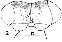
Figure 2. Head of S. biniae.
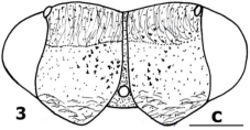
Figure 3. Head of S. flacourtiae.
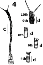
Figure 4. Antenna of S. biniae.
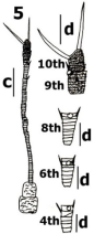
Figure 5. Antenna of S. flacourtiae.
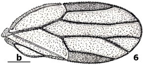
Figure 6. Forewing of S. biniae.
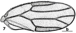
Figure 7. Forewing of S. flacourtiae.
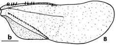
Figure 8. Hindwing of S. biniae.
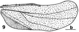
Figure 9. Hindwing of S. flacourtiae.
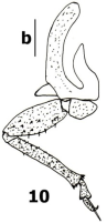
Figure 10. Metathoracic leg of S. biniae.
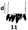
Figure 11. metatibial apical end of S. biniae.
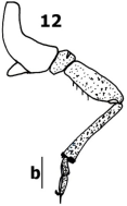
Figure 12. Metathoracic leg of S. flacourtiae.
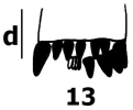
Figure 13. metatibial apical end of S. flacourtiae.
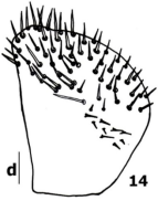
Figure 14. Male proctiger of S. biniae.
Figure 15. Male proctiger of S. flacourtiae.
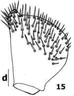
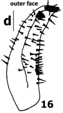
Figure 16. Paramere outer face of S. biniae.
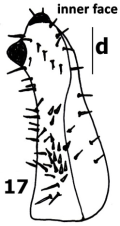
Figure 17. Paramere inner face of S. biniae.
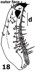
Figure 18. Paramere outer face of S. flacourtiae.
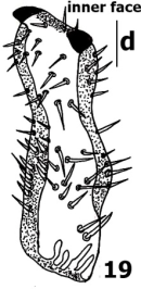
Figure 19. Paramere inner face of S. flacourtiae.
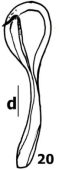
Figure 20. Aedeagus of S. biniae.
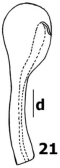
Figure 21. Aedeagus of S. flacourtiae.
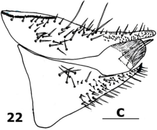
Figure 22. Female genitalia of S. biniae.
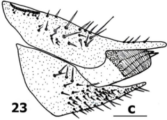
Figure 23. Female genitalia of S. flacourtiae.
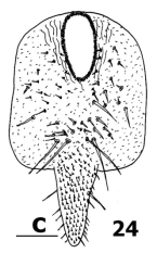
Figure 24. Female proctiger of S. biniae.
Scales: a: 2 mm; b: 0.8 mm; c: 0.4 mm; d: 0.2 mm.

Download: Download full-size image

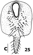
Figure 25. Female proctiger of S. flacourtiae.
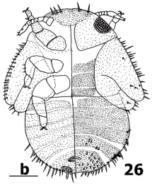
Figure 26. Fifth larval stage, left dorsal view and right ventral view of S. biniae.
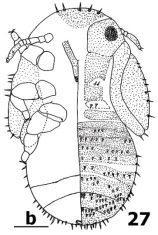
Figure 27. Fifth larval stage, left dorsal view and right ventral view of S. flacourtiae.
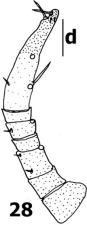
Figure 28. Antenna of S. biniae.
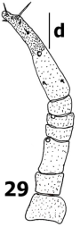
Figure 29. Antenna of S. flacourtiae.
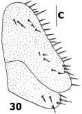
Figure 30. Forewing pad of S. biniae.
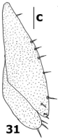
Figure 31. Forewing pad of S. flacourtiae.
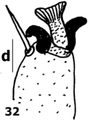
Figure 32. Tarsal arolium of S. biniae.
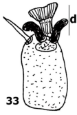
Figure 33. Tarsal arolium of S. flacourtiae.
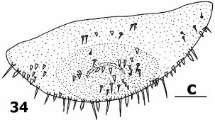
Figure 34. Caudal plate in dorsal view of S. biniae.
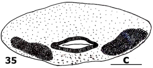
Figure 35. Caudal plate in ventral view of S. biniae. Caudal plate in ventral view of S. biniae.
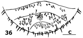
Figure 36. Caudal plate in dorsal view of S. flacourtiae.
Scales: b: 0.8 mm; c: 0.4; d: 0.2 mm.

Download: Download full-size image

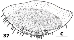
Figure 37. Caudal plate in ventral view of S. flacourtiae.
Table 1. Measurements (mm) and ratios of Syntomoza spp. adults species (N= number of measured specimens).

Parameters

Syntomoza biniae sp. nov. Males

Syntomoza biniae sp. nov. Females

Syntomoza flacourtiae sp. nov. Males

Syntomoza flacourtiae sp. nov. Females

N

Min

Max

Average

N

Min

Max

Average

N

Min

Max

Average

N

Min

Max

Average

BL

40

2.0

3.0

2.34

40

2.0

3.0

2.7

40

2.4

3.0

2.8

40

2.8

3.8

3.35

BW

40

0.80

1.12

0.90

40

0.92

1,2

1.03

40

0.88

1.2

1.04

40

1.12

1.28

1.18

HW

40

0.72

0.88

0.77

40

0.8

1.0

0.86

40

0.68

1.0

0.8

40

0.8

0.92

0.85

AL

40

0.68

1.0

0.83

40

0.68

1.0

0.86

40

1.0

1.12

1.07

40

1.0

1.12

1.08

F1L

40

0.2

0.24

0.22

40

0.2

0.24

0.22

40

0.2

0.2

0.2

40

0.2

0.24

0.22

WL

40

1.6

2.0

1.82

40

1.8

2.8

2.25

40

2.2

2.2

2.2

40

2.4

2.8

2.6

WW

40

0.8

0.92

0.84

40

0.88

1.2

1.04

40

0.88

1.0

0.96

40

1.0

1.28

1.17

wL

40

1.4

2.0

1.60

40

1.6

2.4

1.92

40

1.6

2.0

1.8

40

2.0

2.2

2.18

wW

40

0.52

0.8

0.60

40

0.6

0.8

0.71

40

0.6

0.8

0.69

40

0.8

0.88

0.82

MTL

40

0.4

0.6

0.53

40

0.4

0.68

0.59

40

0.48

0.6

0.54

40

0.52

0.6

0.59

MFL

40

0.36

0.48

0.40

40

0.4

0.52

0.42

40

0.4

0.52

0.48

40

0.4

0.6

0.49

MPL

40

0.2

0.4

0.26

/

/

/

/

40

0.32

0.40

0.36

/

/

/

/

PL

40

0.2

0.28

0.20

/

/

/

/

40

0.24

0.32

0.30

/

/

/

/

FPL

/

/

/

/

40

0.68

1.0

0.79

/

/

/

/

40

0.68

0.8

0.75

PSPL

/

/

/

/

40

0.4

0.6

0.47

/

/

/

/

40

0.4

0.6

0.47

DAL

40

0.2

0.28

0.25

/

/

/

/

40

0.2

0.24

0.21

/

/

/

/

BL/HW

40

2.78

3.40

3.03

40

2.5

3.0

3.13

40

3.52

3.0

3.5

40

3.5

4.13

3.94

BL/BW

40

2.5

2.67

3.78

40

2.17

2.5

2.62

40

2.72

2.5

2.69

40

2.5

2.96

2.83

AL/HW

40

1.11

1.13

1.07

40

0.85

1.0

1.0

40

1.47

1.12

1.33

40

1.25

1.21

1.27

F1/HW

40

0.28

0.27

0.28

40

0.25

0.24

0.25

40

0.29

0.20

0.25

40

0.25

0.26

0.25

AL/F1

40

4.0

4.16

3.77

40

3.4

4.16

3.90

40

5.0

5.6

5.35

40

5.0

4.66

4.90

WL/HW

40

2.22

2.27

2.36

40

2.25

2.8

2.61

40

3.23

2.2

2.75

40

3.0

3.04

3.05

WL/WW

40

2.0

2.17

2.16

40

2.04

2.33

2.16

40

2.5

2.2

2.29

40

2.4

2.18

2.22

WL/wL

40

1.14

1.00

1.13

40

1.12

1.16

1.17

40

1.37

1.1

1.22

40

1.2

1.27

1.19

MTL/HW

40

0,55

0,68

0.68

40

0.55

0.68

0.68

40

0.70

0.60

0.67

40

0.65

0.65

0.69

PL/HW

40

0.27

0.45

0.33

/

/

/

/

40

0.47

0.40

0.45

/

/

/

/

FPL/FSPL

/

/

/

/

40

1.7

1.66

1.68

/

/

/

/

40

1.7

1.33

1.59

LR

40

0.6

0.8

0.60

40

0.6

0.8

0.74

40

0.6

0.8

0.67

40

0.8

1.0

0.86

LM+Cu1

40

0.6

0.8

0.60

40

0.6

0.8

0.74

40

0.4

0.6

0.51

40

0.6

0,92

0,71

LM1+2

40

0.8

1.0

0.86

40

1.0

1.4

1.21

40

1.0

1.2

1.03

40

1.0

1.4

1.18

LM3+4

40

0.6

0.8

0.66

40

0.8

1.2

0.93

40

0.8

0.8

0.8

40

0.8

1.2

0.94

LPt

40

1.2

1.6

1.25

40

1.2

2.0

1.8

40

1.2

1.6

1.36

40

1.2

2.0

1.63

m1

40

0.4

0.6

0.44

40

0.4

0.6

0.44

40

0.6

0.8

0.65

40

0.8

0.88

0.80

Adult legend: BL, body length; BW, body width; HW, head width; AL, antenna length; F1L, length of first antennal flagellomere; WL, forewing length; WW, forewing width; wL, hindwing length; wW, hindwing width; MTL, metatibial length; MFL, metafemur length; MPL, male proctiger length; PL, paramere length; DAL, length of distal segment of aedeagus; FPL, female proctiger length; FSPL, female subgenital plate length; LR, R vein length of forewing; LM1+2, M1+2 vein length of forewing; LM3+4, M3+4 vein length of forewing; LPt, pterostigma length; m1, m1 cell length of forewing.
Table 2. Measurements (mm) and ratio of Syntomoza spp. fifth instar larva (N= number of measured specimens).

Parameters

Syntomoza biniae sp. nov.

Syntomoza flacourtiae sp. nov.

N

Minimum

Maximum

Average

N

Minimum

Maximum

Average

BL

05

1.63

1.81

1.70

04

2.09

2.36

2.22

BW

05

0.81

0.81

0.81

04

0.81

0.90

0.86

AL

05

0.4

0.45

0.42

04

0.58

0.63

0.62

MTL

05

0.67

0.69

0.68

04

0.72

0.72

0.72

WL

05

0.11

0.11

0.11

04

0.11

0.14

0.13

BL/BW

05

2.12

2.62

2.40

04

2.58

2.62

2.58

Fifth instar larva legend: BL, body length; BW, body width; AL, antenna length; WL, forewing-pad length; MTL, metatibial length.
4. Discussion
Syntomoza biniae sp. nov. differs from others Syntomoza species in the general colour and wing pattern. In S. magna , membrane of forewings pale yellowish brown to dirty yellowish brown, scattered with numerous irregular dark brown spots, and sometimes dark patches; body colouring in general dirty yellowish brown to ochreous, scattered with numerous dark brown to black spots and several dark patches on head and thorax. In Anomoterga tatuata / Syntomoza tatuata , general colour dark brown to black with lighter brown markings and stripes on the thorax and abdomen. In S. unicolor , dorsal surface of head and body evenly brown, ventral surface ochreous; wings transparent, whitish, veins light ochreous.
Head in Syntomoza biniae sp. nov., with width 0.8-1.12 mm in males and 0.8-1.0 mm in females, vertex sub-rectangular without foveae. It differs from Anomoterga tatuata and Syntomoza tatuata with width of head, less broad, 0.6-0.7 mm, vertex with two foveae on each side of the median suture and situated toward the upper part. It also differs from S. unicolor with width of head, less broad, 0.59-0.67 mm, vertex with fine microsculpture.
Antenna in Syntomoza biniae sp. nov., longer than the width of the head; basal two segments robust, cylindrical, the third segment is the longest, terminal segment with two almost equal long simple setae at apex, two small simple setae present near each rhinaria on segment 4, 6 and 9. It differs from S. magna with antenna shorter than width of head and it also differs from A. tahuata with first and second antennal segments small and slender. It is close to S. unicolor with antenna bears a pair of short setae on segments 4, 6 and 9, with subequal terminal setae, both longer than segment 10.
Forewing in Syntomoza biniae sp.n membranous and covered with dense spinule surfaces occupying all the surfaces of the wing cells, long and broadest medially; RS vein subparallel with M vein and straights, apex portion of RS flexed downward near apex; cu1 cell very large and longer than the pterostigma. It is close to S. africana and S. unicolor in forewings shape, however, surface spinules cover just the half posterior in C+Sc cell in S. Africana . It differs from S. magna , with forewing somewhat parallelogram shaped, broad, cell m2 apparently smaller than cu1.
Hindwing in Syntomoza biniae sp. nov. long and narrowed, narrowly rounded and upturned apically, the membrane beset numerous punctations, only R+M+Cu1 vein conspicuous, costal margin armed with 2+7 ungrouped setae basally, two setae before costal break and seven other after costal break and 1 hooked seta apically (hamulus). It differs from A. tatuata , with the obscure venation developed in the form of fine ridges along which the punctations are arranged in more or less definite single, double, and irregular rows. It also differs from S. magna with all vein conspicuous, costal margin armed with 3+3 ungrouped setae basally, membrane subopaque.
Metacoxa in Syntomoza biniae sp. nov., with very developed meracanthus, laterally straight, horn shaped; metatibia enlarged at its apical end and bearing an open crown of 10 sclerotized black apical spurs and a group of 6 small teeth at the apex. It differs from A. tatuata , with 8-10 sclerotized black apical spurs and 2-4 small teeth at the apex; 10 spurs and 3 small stout setae in S. Africana .
Male proctiger in Syntomoza biniae sp. nov. simple, broad and short; in profile parallel-margined, broad basally, anterior margin straight, lateral lobe large, broadly rounded in the middle, narrowed apically, obliquely truncate at apex bearing a row of long simple setae; bearing on apical two-thirds surface, dense moderately long setae. It differs from A. tatuata with apical part longest, narrowed, incurved on anterior margin apically, horizontally truncate at apex without a row of long setae. It differs from S. africana and S. unicolor with anterior margin slightly sinuate, lateral lobe large, broadly truncated in the middle narrowed apically, incurved on posterior margin apically, obliquely truncate at apex. It also differs from S. magna with anterior margin straight in half basally, slightly incurved in half apically, lateral lobe large, prominent subtriangular broadly rounded posteriorly in the middle, horizontally truncate at apex.
Paramere in Syntomoza biniae sp. nov. is almost lamellar, posterior margin slightly curved, broad at base, apical region narrow curved forward bearing two short black thick spine oriented inwards; inner face with a conspicuous fold anteriorly extending obliquely toward apex; mesal surface bears about twenty short and stout setae. It differs from A. tatuta lamellar with a forward directed single claw-like process on the mesal side of the apex. It differs from S. africana with paramere in lateral view, lamellar, narrowed in the middle; apical margin sclerotized, with a forward directed single claw-like process on the mesal side of the apex, inner face with a cluster of seventeen stout setae pointing upward in its basal half. It also differs from S. unicolor with paramere obliquely truncate with each an anterior and posterior sclerotised tooth apically, anterior and posterior margins incurved medially and equally differs from S. magna with paramere long, narrowed to apices, tips pointed and directed cephalad.
Distal segment of aedeagus in Syntomoza biniae sp. nov. almost curved in basal half, dorsal margin weakly sinuate, without expansion basally, with a simple, ovate (spoon-like structure) apical dilation, ductus ejaculatorius relatively long and straight. It differs from S. africana with distal portion of aedeagus relatively short with rounded apical dilatation, ductus ejaculatorius relatively long and sinuate. It differs from A. tatuata , distal segment of aedeagus, with long lateral lobe, apex strongly dilated; it also differs from S. unicolor , with basal portion of aedeagus with transverse folds apically on the inner side; and it equally differs from S. magna with apex enlarged on upper side, dorsal margin strongly curved.
Female subgenital plate in Syntomoza biniae sp. nov. in lateral view subtriangular, very enlarged at the base with distinct hump, strongly narrowed at the apex, with posterior half bent upward and acutely pointed at apex, ventral margin straight upwards. It differs from S. unicolor and S. magna with female subgenital plate in lateral view enlarged at the base, rounded at base, ventral margin truncated upwards.
In Syntomoza biniae sp. nov., circumanal ring elongated oval shape consisting of two rows of pores surrounded by small setae, the outer row circular and the inner one oblongate pores. Dorsal and ventral valvulae slightly curved dorsally, inner valve is shorter than the ventral one, ovipositor acutely pointed. It differs from A. tatuata with the circumanal ring developed as a single row of simple, elongate pores; it also differs from S. unicolor and S. magna , with dorsal and ventral valvulae strongly curved ventrally, ovipositor very long acutely pointed.
For the fifth instar larvae. In Syntomoza biniae sp.n, antenna with a rhinaria formula of 3577 like in S. unicolor .
In Syntomoza biniae sp.n., antennal segments with following numbers of pointed setae: 1(0), 2(0), 3(2), 4(0), 5(1), 6(0), 7(2), seventh segment longest, not differentiated into segments, bearing two equal and slender terminal spines. It differs from S. unicolor with following numbers of pointed setae: 1(3), 2(1), 3(2), 4(0), 5(2), 6(0), 7(1), seventh segment bearing two inequal and slender terminal spines.
Forewing pad in Syntomoza biniae sp.n., with a row of 21 marginal very long lanceolate setae like in S. unicolor ; hindwing pad with four marginal long lanceolate setae similar to S. unicolor .
In Syntomoza biniae sp.n., dorsum of caudal plate with strong micro sculpture consisting of scarcely arranged small sharp cuticular teeth. It differs from S. unicolor with dorsum of caudal plate, with several rows of cuticular teeth mixed to small simple setae.
Anal opening ventral in Syntomoza biniae sp.n., surrounded by only outer circumanal pore ring consisting of a single row of elongated pores; extra pore fields forming continuous inner band, consisting of elongate patches. It differs from S. unicolor with additional inner circumanal pore ring consisting of row of circular pores.
In adults, Syntomoza flacourtiae sp. nov., is close to Syntomoza biniae sp. nov. which is sheltered by the same host plant, nevertheless we note some differences: dark yellow coloration in Syntomoza biniae sp. nov. and dark brown in Syntomoza flacourtiae sp. nov.; specimens relatively larger than one; genal cones non separated from vertex, with fine microsculpture, the anterior margin without setae; the type of venation on the hindwing with all veins conspicuous is different as well as the number of spiny setae on costal margin armed with 2+6 ungrouped instead of 2+7 ungrouped setae basally; metatibia bearing an open crown of 10 sclerotized black apical spurs and a group of 4 small teeth at the apex instead of 10 sclerotized black apical spurs and a group of 6 small teeth; male proctiger in profile medially with well-developed, broadly rounded posterior expansions, rather with two pits, one at base and other at apex, narrowed apically, apical margin rounded; paramere is rather with bulging posterior margin on one-third basally, anterior margin incurved, inner face covered with two row of eight stouter setae situated along anterior margin medially; the shape of subgenital plate in lateral view subtriangular, rounded at the base, ventral margin straight upwards, with an blunt apex, without distinct hump; circumanal ring oval to pear shaped; arrangement of setae on the female proctiger, especially in their middle is denser with a transverse row of very long simple setae instead of two rows as in Syntomoza biniae sp. nov.
In fifth instar larvae: Syntomoza flacourtiae sp. nov. differs from Syntomoza biniae sp. nov. in the form; antenna segments 3 and 5 without numbers of pointed setae, seventh segment bearing two inequal and slender terminal spines; forewing pad with a row of seven marginal long lanceolate setae instead of 21; abdomen on all dorsum surface bearing lanceolate setae and additional like-scaly setae; anus is rather terminal, without additional pore fields.
5. Conclusion
Host plant, Flacourtia indica belonging to Flacourtiaceae family, shelters two news species belonging to Syntomoza genus (Hemiptera: Liviidae). Syntomoza biniae sp. nov., is morphologically different from others species of Syntomoza genus. Adults, has vertex sub-rectangular without foveae; hindwing with membrane beset numerous punctations and only R+M+Cu1 vein conspicuous; male proctiger in profile broad basally, anterior margin straight, lateral lobe large, broadly rounded in the middle, narrowed apically, obliquely truncate at apex bearing a row of long simple setae; paramere with posterior margin slightly curved, broad at base, apical region narrow curved forward bearing two short black thick spine oriented inwards; inner face with a conspicuous fold anteriorly extending obliquely toward apex; mesal surface bears about twenty short and stout setae; distal segment of aedeagus almost curved in basal half, dorsal margin weakly sinuate, without expansion basally, ductus ejaculatorius relatively long and straight; female subgenital plate in lateral view subtriangular, very enlarged at the base with distinct hump, posterior half bent upward and acutely pointed at apex, ventral margin straight upwards; circumanal ring elongated oval shape consisting of two rows of pores, dorsal and ventral valvulae slightly curved dorsally. Fifth instar larvae, with antennal segments with following numbers of pointed setae: 1(0), 2(0), 3(2), 4(0), 5(1), 6(0), 7(2), seventh segment bearing two equal and slender terminal spines; dorsum of caudal plate bearing scarcely arranged small sharp cuticular teeth; anus ventral, surrounded by only outer circumanal pore ring.
Syntomoza flacourtiae sp. nov., is morphologically different from others species of Syntomoza genus and Syntomoza biniae sp. nov. with specific characters: male proctiger in profile medially with well-developed, broadly rounded posterior expansions, rather with two pits, one at base and other at apex, narrowed apically, apical margin rounded; paramere is rather with bulging posterior margin on one-third basally, anterior margin incurved, inner face covered with two row of eight stouter setae situated along anterior margin medially; circumanal ring oval to pear shaped; arrangement of setae on the female proctiger, especially in their middle is denser with a transverse row of very long simple setae. In fifth instar larvae: antenna segments 3 and 5 without numbers of pointed setae; forewing pad with a row of seven marginal long lanceolate; abdomen on all dorsum surface bearing lanceolate setae and additional like-scaly setae; anus terminal, without additional pore fields.
This study enriched the biodiversity of jumping plant louse of Syntomoza genus and also the pests of African medicinal plants to survey.
Abbreviations

LZUY

Laboratory of Zoology, Higher Teachers’ Training College, University of Yaounde I, Cameroon

Acknowledgments
We sincerely thank Laboratory of Zoology of the Higher Teachers’ Training College of the University of Yaounde I for its best work conditions and collections conservation. We also thank Pr YANA Wenceslas for the preliminary identification of the psyllids species of the collection from Adamawa Region.
Author Contributions
Louis Djakbe Dayang: Conceptualization, Data curation, Formal Analysis, Investigation, Methodology, Resources, Writing – original draft, Writing – review & editing
Joseph Lebel Tamesse: Formal Analysis, Methodology, Supervision, Validation, Writing – review & editing
Funding
This work is not supported by any external funding source.
Data Availability Statement
The data supporting the outcome of this research work has been reported in this manuscript.
Conflicts of Interest
The authors declare no conflicts of interest.
References
[1] Burckhardt D. Biology, ecology, and evolution of gall-inducing Psyllids (Hemiptera: Psylloidea). In: Raman R., Schaefer C. W. & Withers T. M. (eds.): Biology, ecology, and evolution of gallinducing arthropods. Science Publishers, Enfield, Plymouth, 2005, xxi, 143-157.
[2] Hollis D. Australian Psylloidea: jumping plantlice and lerp insects. Australian Biological Ressources Study, Canberra. 2004, xvi + 216.
[3] Hodkinson I. D. Life cycle variation and adaptation in jumping plant lice (Insecta: Hemiptera: Psylloidea): a global synthesis, Journal of Natural History. 2009, 43(1-2), 65-179.
[4] Burckhardt D., Ouvrard D., Queiroz D and Percy D. Psyllid host-plants (Hemiptera: Psylloidea): resolving a semantic problem, Florida Entomologist. 2014, 97(1), 242-246.
[5] Burckhardt D. Psylloid pests of temperate and subtropical crops and ornamental plants (Hemiptera, Psyllidea): a review, Trends in Agricultural Sciences, Entomology. 1994, 2, 173-186.
[6] Percy D. M. Psyllids or «jumping plant lice» (Psylloidea, Hemiptera), University of British Columbia, Department of Botany, Beaty Biodiversity Museum. 2002.
[7] Crawford, D. L. New genera and species of psyllidae from the Philippine Islands, The Philippin. Journal of Science. 1914, 8, 293-301, 1 plate.
[8] Burckhardt, D., Ouvrard, D. and Percy, D. M. An updated classification of the jumping plant-lice (Hemiptera: Psylloidea) integrating molecular and morphological evidence, European Journal of Taxonomy. 2021, 736, 137-182.
[9] Burckhardt D. & Misfud D. Jumping plant-lice of the Paurocephalinae (Insecta, Hemiptera, Psylloidea). Systematics and phylogeny, Contibutions to Natural History (Bern). 20032, 3-34.
[10] Burckhardt, D., D. C. Alene, D. Ouvrard, J. L. Tamesse and J. Messi, 2006. Afrotropical members of the jumping plant-louse genus Diclidophlebia (Hemiptera: Psylloidea). Invert. Syst., 20: 367-393.
[11] Vondracek, K. Jumping plant-lice (Psylloidea-Homoptera) of Central Africa: Part I (Congo), Sb. Entomol. Odd. Ndr. Mus. Praze. 1963, 35, 263-290.
[12] Messi, J., D. C. Alene and J. L. Tamesse. Diclidophlebia xuani (Homoptera-Psylloidea) new species of psyllid associated with Ricinodendron heudelotii, Ann. Fac. Sci. Univ. Yaounde I. Ser. Sci. Vie Nat. 1998, 34, 233-237.
[13] Tamesse J. L. and Dayang L. D. Newly described psyllid Diclidophlebia andjigae sp.n. (Hemiptera: Liviidae), on Grewia venusta (Tiliaceae) from Cameroon, Journal of Entomolology. 2018, 15, 19-17.
[14] Yana, W., Dzokou V. J., Ndankeu Mveyo Y. P. and Tamesse J. L. Two species of psyllids genus Paurocephala (Hemiptera: Psyllidae) pest insects associated to Coronnaceae in Cameroon, Entomology Journals. 2019, 4(2), 13-19.
[15] Dayang L. D., Dzokou V. J. and Tamesse J. L. Paurocephala famendongoeis sp. n. (Hemiptera: Psyllidae), Insect Pest of Psorospermum febrifugum (Hypericaceae) from Adamawa-Cameroon, American Journal of Zoology. 2023, 6(1), 1-8.
[16] Mifsud D. & Burckhardt D. Taxonomy and phylogeny of the old world jumping plant-louse genus Paurocephala (Insecta, Hemiptera, Psylloidea), Journal of Natural History. 2002, 36, 1887-1986.
[17] Bonnet P., Arbonnier M. and Grard P., “Woody plants of the Sahel. Graphic identification tool” V. 1.0 CIRAD. 2008.
[18] Adjanohoun E. J., Ake Assi L., Floret J. J., Koumare M., Ahyi M. R. A., Raynal J. Contribution to ethnobotanical and floristic studies in Mali. Paris, Agency for Cultural and Technical Cooperation (Actc). 1985, 56.
[19] Ouôba P, Lykke AM, Boussim J, Guinko S. The medicinal flora of the Niangoloko Classified Forest (Burkina Faso). Studies on the flora and vegetation of Burkina Faso and neighboring countries. 2006, Vol. 10. Editions Verlag Natur & Wissenschaft, Solingen: Francfort and Ouagadougou; 5-12.
[20] Jin Hyung Kwon and Yong Jung Kwon. Insect Fauna of Korea, Psylloidea, Arthropoda: Insecta: Hemiptera: Sternorrhyncha, National Institute of Biological Resources, Ministry of Environment. 2020, 9(9), 405.
[21] Klyver F. D. Anomoterga tatuata, new genus and new species and other chermidae from the Marquesas, Pacific Entomological Survey Publication I. 1932, 8, 1-3.
[22] Martoni F. and Brown S. D. J. An annotated checklist of the Cook Islands psyllids with keys to the species and two new records (Hemiptera, Psylloidea), ZooKeys. 2018, 811, 91-108.
[23] D. Burckhardt and P. Lauterer. The jumping plant-lice of Iran (Homoptera, Psylloidea), “Swiss Journal of Zoology”. 1993, 100(4), 829-898.
Cite This Article
  • APA Style

    Dayang, L. D., Tamesse, J. L. (2026). Two News Species of Psyllids Genus Syntomoza (Hemiptera: Liviidae) Pest Insects Associated with Flacourtia Indica (Flacourtiaceae) in Cameroon. American Journal of Zoology, 9(1), 8-22. https://doi.org/10.11648/j.ajz.20260901.12

    Copy | Download

    ACS Style

    Dayang, L. D.; Tamesse, J. L. Two News Species of Psyllids Genus Syntomoza (Hemiptera: Liviidae) Pest Insects Associated with Flacourtia Indica (Flacourtiaceae) in Cameroon. Am. J. Zool. 2026, 9(1), 8-22. doi: 10.11648/j.ajz.20260901.12

    Copy | Download

    AMA Style

    Dayang LD, Tamesse JL. Two News Species of Psyllids Genus Syntomoza (Hemiptera: Liviidae) Pest Insects Associated with Flacourtia Indica (Flacourtiaceae) in Cameroon. Am J Zool. 2026;9(1):8-22. doi: 10.11648/j.ajz.20260901.12

    Copy | Download

  • @article{10.11648/j.ajz.20260901.12,
      author = {Louis Djakbe Dayang and Joseph Lebel Tamesse},
      title = {Two News Species of Psyllids Genus Syntomoza (Hemiptera: Liviidae) Pest Insects Associated with Flacourtia Indica (Flacourtiaceae) in Cameroon},
      journal = {American Journal of Zoology},
      volume = {9},
      number = {1},
      pages = {8-22},
      doi = {10.11648/j.ajz.20260901.12},
      url = {https://doi.org/10.11648/j.ajz.20260901.12},
      eprint = {https://article.sciencepublishinggroup.com/pdf/10.11648.j.ajz.20260901.12},
      abstract = {Background: Psyllids mainly through their larvae which suck sap from their host plants can be harmful by causing the transmission of diseases through their saliva. Objective: The aim of this study was to describe the news species of psyllids genus Syntomoza pest insects associated with Flacourtia indica (Flacourtiaceae). Methods: Psyllids specimens were collected using an entomological net and a mouth aspirator. The specimens were preserved in 70% ethanol, mounted on slides. Identifications, measurements and illustrations were made using a Microscope. Results: Syntomoza biniae sp. nov., have vertex sub-rectangular without foveae; male proctiger in profile, with lateral lobe large, broadly rounded in the middle, narrowed apically, obliquely truncate at apex; paramere with apical region narrow curved forward bearing two short black thick spine oriented inwards; inner face with a conspicuous fold anteriorly extending obliquely toward apex; distal segment of aedeagus almost curved in basal half, dorsal margin weakly sinuate; female subgenital plate in lateral view subtriangular, very enlarged at the base with distinct hump; dorsal and ventral valvulae slightly curved dorsally. Fifth instar larvae, with antennal segments with following numbers of pointed setae: 1(0), 2(0), 3(2), 4(0), 5(1), 6(0), 7(2); dorsum of caudal plate bearing scarcely arranged small sharp cuticular teeth; anus ventral, surrounded by only outer circumanal pore ring. Syntomoza flacourtiae sp. nov., with male proctiger in profile medially with well-developed, broadly rounded posterior expansions, rather with two pits, one at base and other at apex, narrowed apically, apical margin rounded; paramere is rather with bulging posterior margin on one-third basally, anterior margin incurved, inner face covered with two row of eight stouter setae situated along anterior margin medially; circumanal ring oval to pear shaped. In fifth instar larvae: antenna segments 3 and 5 without numbers of pointed setae; forewing pad with a row of seven marginal long lanceolate; abdomen on all dorsum surface bearing lanceolate setae and additional like-scaly setae; anus terminal, without additional pore fields. Damage caused by Syntomoza spp. includes distortion and winding of the lateral edges of young leaves. Conclusion: Taxonomic studies indicated that, Syntomoza biniae sp. nov. and Syntomoza flacourtiae sp. nov. are the news species of Liviidae family described from Adamawa Region of Cameroon.},
     year = {2026}
    }
    

    Copy | Download

  • TY  - JOUR
    T1  - Two News Species of Psyllids Genus Syntomoza (Hemiptera: Liviidae) Pest Insects Associated with Flacourtia Indica (Flacourtiaceae) in Cameroon
    AU  - Louis Djakbe Dayang
    AU  - Joseph Lebel Tamesse
    Y1  - 2026/02/04
    PY  - 2026
    N1  - https://doi.org/10.11648/j.ajz.20260901.12
    DO  - 10.11648/j.ajz.20260901.12
    T2  - American Journal of Zoology
    JF  - American Journal of Zoology
    JO  - American Journal of Zoology
    SP  - 8
    EP  - 22
    PB  - Science Publishing Group
    SN  - 2994-7413
    UR  - https://doi.org/10.11648/j.ajz.20260901.12
    AB  - Background: Psyllids mainly through their larvae which suck sap from their host plants can be harmful by causing the transmission of diseases through their saliva. Objective: The aim of this study was to describe the news species of psyllids genus Syntomoza pest insects associated with Flacourtia indica (Flacourtiaceae). Methods: Psyllids specimens were collected using an entomological net and a mouth aspirator. The specimens were preserved in 70% ethanol, mounted on slides. Identifications, measurements and illustrations were made using a Microscope. Results: Syntomoza biniae sp. nov., have vertex sub-rectangular without foveae; male proctiger in profile, with lateral lobe large, broadly rounded in the middle, narrowed apically, obliquely truncate at apex; paramere with apical region narrow curved forward bearing two short black thick spine oriented inwards; inner face with a conspicuous fold anteriorly extending obliquely toward apex; distal segment of aedeagus almost curved in basal half, dorsal margin weakly sinuate; female subgenital plate in lateral view subtriangular, very enlarged at the base with distinct hump; dorsal and ventral valvulae slightly curved dorsally. Fifth instar larvae, with antennal segments with following numbers of pointed setae: 1(0), 2(0), 3(2), 4(0), 5(1), 6(0), 7(2); dorsum of caudal plate bearing scarcely arranged small sharp cuticular teeth; anus ventral, surrounded by only outer circumanal pore ring. Syntomoza flacourtiae sp. nov., with male proctiger in profile medially with well-developed, broadly rounded posterior expansions, rather with two pits, one at base and other at apex, narrowed apically, apical margin rounded; paramere is rather with bulging posterior margin on one-third basally, anterior margin incurved, inner face covered with two row of eight stouter setae situated along anterior margin medially; circumanal ring oval to pear shaped. In fifth instar larvae: antenna segments 3 and 5 without numbers of pointed setae; forewing pad with a row of seven marginal long lanceolate; abdomen on all dorsum surface bearing lanceolate setae and additional like-scaly setae; anus terminal, without additional pore fields. Damage caused by Syntomoza spp. includes distortion and winding of the lateral edges of young leaves. Conclusion: Taxonomic studies indicated that, Syntomoza biniae sp. nov. and Syntomoza flacourtiae sp. nov. are the news species of Liviidae family described from Adamawa Region of Cameroon.
    VL  - 9
    IS  - 1
    ER  - 

    Copy | Download

Author Information